An Overview of the Chemistry of Color Vision

Intro
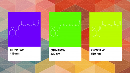
The human eye contains two different types of cells, rods and cones. Rods are used for night vision, and cones enable color vision. Further, there are three types of cones; short, medium, and long-wavelength cones. They are named this way because each of them best absorb photons of light of three different wavelengths of light, corresponding to the colors in the picture above. Specific proteins, called opsins, bound to retinal--a chromophore--determine which color the cone or rod will absorb.
-3,7-Dimethyl-9-(2,6,6-trimethylcyclohexen-1-yl)nona-2,4,6,8-tetraenal)")
Retinal
Retinal is a derivative of Vitamin A, and can be found bound to either Rhodopsin, the photopigment in rod cells for night vision, or the three color vision photopigments in their respective cone cells. Retinal absorbs photons of lights which causes a change in the structure of the molecule. This change then leads to a conformational change in the protein bound to the retinal, triggering a cascade of chemical events which ends in a nerve signal being sent to the brain. All of this happens in a a matter of picoseconds.

Opsns: The Proteins that Bind Retinal
The proteins that bind retinal are found in the membranes of rod cone cells, where they can most efficiently absorb photons of light. They are called opsins, and they belong to a special type of class of proteins known as G-proteins. G-proteins are membrane-bound proteins with a structure consisting of seven alpha-helices. The genes that code for these color vision photopigments are labeled in the image at the beginning of this article.


