Keratoacanthoma – Symptoms, Causes, Treatment, Pictures

Keratoacanthomas refer to benign but fast-growing bumps particularly on the sun-exposed areas. They are described as dome-like bumps with open craters at the center and are quite visible on the arms, legs, head or torso. These bumps rarely appear in multiples or spread further. They are usually painless but are extremely itchy. Their presence can cause a great deal of concern especially if they are found on the face. They can also affect the optimum functioning of the affected area.
The lesions caused by keratoacanthomas may resolve even without treatment. But doctors and patients share the same sentiments with regards to the presence of these bumps due to their resemblance to a form of skin cancer called squamous cell carcinoma. Treatment often involves surgical removal of the bumps either through curettage, freezing, excision and radiotherapy.
What causes Keratoacanthomas?
Medical experts do not exactly know why and how keratoachanthoma develops. But extensive studies showed that the blisters develop on the area where skin usually grows and exposed to sunlight. Moreover, it had also been discovered that the blisters form on the areas that had sustained a minor injury, whether the affected person is aware of it or not. The cells start to proliferate and cause a mass to grow in the hair follicle. Other factors linked by experts to the development of keratoacanthomas are:
- Exposure to hazardous chemicals or substances at work
- HPV (human paplillomavirus) infection
- Direct trauma
Keratoacanthoma can come in different forms, and this can be:
- Giant
- Appear in multiples
- Appear in multiples in a localized area
- Appear all over the body
- Solitary but grows aggressively
Who are at risk of developing Keratoacanthomas?
Some individuals are more susceptible to developing keratoacanthomas than others because they have the risk factors, such as:
- Working in an industry that constantly exposes them to harsh chemicals and substances.
- Staying under the sun for long periods of time.
- Having compromised immunity or suffering from an HPV infection.
- Sustaining direct and/or minor trauma on the skin.
- Being male, light-skinned and older than 50 years old.
Research showed that keratoacanthomas occur more frequently in males than females. It has been found to affect approximately 1 out of 1,000 individuals. The condition is very much prevalent in older, lighter complexioned people who are between 50 and 64 years old. On the onther hand, it rarely affects young adults and dark-skinned individuals.
What does Keratoacanthomas look like?
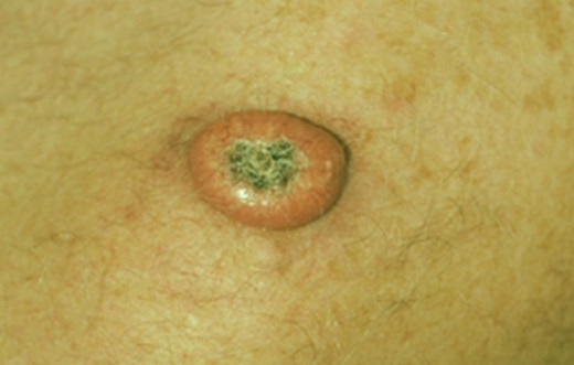
Keratoacanthoma initially appears as a tiny bump with a reddish or flesh hue. It eventually develops into a dome-shaped bump with an open crater at the center in four weeks time. White crusty substance, which is composed of keratin, is visible on the open crater and cause horn-like projections. Keratoacanthomas does not usually grow less than 5 cm in diameter but slowly decreases in size from 6 weeks onwards, leaving a depressed scar at the center.
Keratoacanthomas are notably visible on the ears, face, forearms, scalp, neck, back of hands and lower legs. The bump is painless but itchy and usually grows solitary. However, some individuals develop multiple bumps which can be as big as 15cm or spread to other parts of the body.
Why seek treatment for Keratoacanthomas?
Due to its resemblance to the skin cancer known as squamous cell carcinoma, doctors advise patients to seek treatment. Still, although it may get bigger and bigger if left untreated, keratoacanthomas is different from skin cancer because it self-destructs and resolve over time even without treatment. However, this may take long, from 6 months to 1 year.
Before diagnosing keratoacanthoma, the doctor will first discount the possibility of serious skin cancer involving dome-shaped bumps, such as squamous and basal cell carcinoma as well as actinic and seborrhoeic keratosis. He or she will get a sample of the tissue from the bump for laboratory analysis. Once serious skin cancer is completely ruled out, the doctor will confirm the keratoacanthoma diagnosis by conducting a skin biopsy. After which, he or she will plan the most appropriate treatment method for the patient, taking into consideration the location and size of the bump.
What are the various treatment options for Keratoacanthomas?
Treatment for keratoacanthoma is aimed towards removing the bump. This may require:
- Curettage
- Freezing
- Excision
- Scrape-and-burn
- Radiation therapy
- Mohs micrographic surgery
The doctor will be the one to determine which surgical procedure is best for the patient depending on the size, position and location of the growths. Treating keratoacanthomas with medicines is possible but this is only reserved for cases where surgical intervention is not applicable or multiple or generalized growths are involved. Moreover, antimicrobial ointments or creams provide relief to irritated and inflamed tissues nearby.
The patient should follow the doctor’s advice to wear good sunscreen protection after treatment to prevent further damage caused by harmful UV rays. He or she should also visit the doctor regularly to ensure that new growths have not developed.
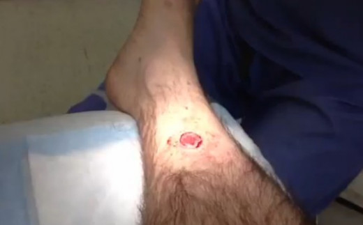
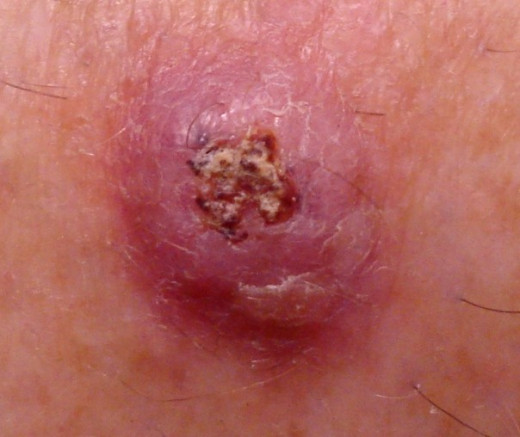
Keratoacanthoma Pictures