What is slit lamp microscopy?

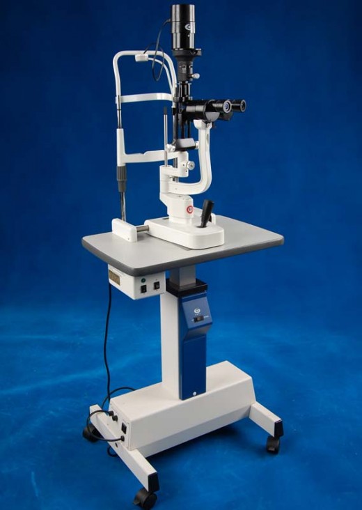
Slit lamp microscopy is a type of eye evaluation that is done utilizing a microscope. This microscopic eye exam is primarily done to reveal foreign objects or deformities in the front part of the eyes but an ophthalmologist can also use this exam to evaluate the rear parts of the eyes. A slit lamp microscope is a very low powered microscope providing a bright light source, which shines a light layer over the eye and then this ophthalmological device is used at the same time to carry out the review on other parts of the eye.
The slit lamp microscope also helps the optometrist to evaluate three dimensions in the various sections of the eye. Clear cornea or outer covering of the eye is visible during the test, and also the doctor is able to visualize parts of eye including lens, iris, and vitreous gel, which is the liquid occupying a good deal of space in the middle section of the eye. In some patients with severe eye problems, the ophthalmologist can also decide to place a special lens between the lamp and the cornea or can put it directly on top of the cornea to allow a deep evaluation of eye structures including optic nerve, retina, and the drainage angle.
In order to perform slit lamp microscopy, the patient needs to sit on a chair and the optometrist directs the slit lamp device in front of his eyes. Just like doing some visual field testing, the rest of patient’s forehead and chin will support against the slit lamp equipment.
The doctor then administers a fluorescein dye (orange color dye) into the defective eye in order to easily detect problems such as infection, injury, or foreign bodies. Once the slit lamp microscopy is done in the front parts of the eye, the doctor carefully inserts dilating eye drops in both eyes to allow full dilation of pupils. Once the eyes are fully dilated, the slit lamp microscopy is again performed on the eyes. This time the examination is done in the back parts of the eyes.
A doctor as part of a routine eye exam can perform slit lamp microscopy on a frequent basis. Also, this exam can be done for patients with eye disorders and also in case of patients reporting blurry vision, foreign body sensation, chemical contacts, or workplace eye injuries like sheet metal getting into the eyes.
The test is performed in conjunction with other things including eye tests and tonometry. This microscope guided eye exam can also be performed on patients who have problems in front parts of the eyes such as iritis, cataracts, conjunctivitis, beginning of any eye infection, or corneal damage. Complete evaluation of eyes with slit lamp microscopy is also beneficial for patients having eye diseases like glaucoma and/or macular degeneration.


