A Description of the Structure and Function of the Cells and Tissues of a Typical Higher Plant

Higher plants are the angiosperms, gymnosperms, ferns and clubmosses etc. These tracheophytes are classified as such because they have lignified tissues to enable them to conduct photosynthetic products, water and minerals through the plant. Thus a ‘typical higher plant’ could be any particular plant in these categories (Esau, 1953).
Figure 1 Diagram of a True Leaf
Click thumbnail to view full-size
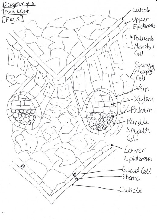
Figure 2 Diagram of Xylem
Click thumbnail to view full-size
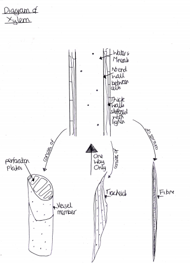
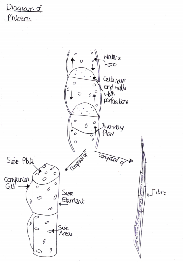
Figure 3 Diagram of Phloem
Click thumbnail to view full-size
The Three Major Tissue Types and Their Constituent Parts
The three major tissue types of plants are the dermal, ground and vascular tissues. Their constituent parts will be explained further.
The first of the major tissue types is that of dermal [Fig 1]. It covers the outer surface of herbaceous plants. This tissue type is composed of epidermal and peridermal cells. (Purves et al, 2004).
Epidermal cells are closely packed cells with little intercellular space that secrete a waxy cuticle that helps to prevent water loss and provide a barrier against pathogens, insects and fungi. The cuticle is transparent to allow light to go through and this is achieved by epidermal cells having no chloroplasts and so do not contain the green pigment chlorophyll. They also do not have a nucleus usually. They can sometimes contain epidermal hairs. These hairs slow the plant’s water loss by decreasing the flow of air over the surface of the plant (Pearson Education, 2011). The cell types which compose the epidermis are: trichrome (hair), subsidiary, epidermal and guard. These guard cells cover the stomata which are small openings or pores, in the plant to allow gas exchange for photosynthesis and respiration. Thus, these cells help to regulate the exchange of oxygen, carbon dioxide and water vapour through the stomata.
Peridermal cells create secondary tissues developed from cork cambrium. It consists of phellem (which are dead cells), phellogen (cork cambrium) and phelloderm. These peridermal cells provide insulation for the plant by covering the outside of roots and stems. They also prevent excessive water loss, protect the plant against pathogens and injury. When they are present, they completely replace the epidermis.
Another main tissue type is vascular tissue. Its function is to transport food, water, hormones and minerals within the plant. The cell types it is composed of are xylem, phloem, parenchyma and cambium.
The cell types in xylem are vessel member, tracheid, fibre and parenchyma [Fig 2]. Tracheids are the more primitive of the cells. They have long, tapered end-plates that connect the cells to each other. Vessel elements, however, are shorter, wider and have no end plates. They are only found in angiosperms, the most evolved group of plants. Xylem cells have woody, lignin-impregnated walls. They conduct the water and minerals from the roots to the leaves and where ever they are required.
Phloem is a living tissue that carries sucrose and other sugars from where they are produced during photosynthesis to where they are required in the plant [Fig 3]. This process is called translocation. They are located on the outside of the xylem. They consist of: sieve tube members, sieve cells (which the food flows through), companion cells (which retain their nucleus to control the sieve cells), fibre, sclerid and parenchyma cells.
The cambium (and in particular the procambium) is the meristematic tissue that produces the primary vascular tissues (xylem and phloem). It develop directly beneath the growing tip, right next to the new leaf primordial.
Ground cells are another main tissue type. It comprises of the bulk of the primary plant body. The most common types are parenchyma, collenchyma and sclerenchyma.
Collenchyma cells provide plants with support. They have a thick call wall but they are not as strong as lignin because it is dead while collenchyma cells are alive at maturity however this does mean that they are more flexible (Cutler, 2005). They occur as part of the vascular bundles or on the corners of angular stems.
Sclerenchyma cells also support the plant however they are dead at maturity. They have thickenings in their secondary walls and are comprised of cellulose, hemicellulose and lignin. Some are isolated in soft tissues e.g. in pears, which gives them their gritty texture. There are two types of sclerenchyma cells, those of sclereids and fibres. Fibre cells have thick walls and small lumen. They consist of a pointed shape and simple pits. Sclereids, however, have an irregular shape which is maintained with thick lignified cell walls and so a very small lumen. They also have simple pits, called canals, to link adjacent cells through the cell wall.
The parenchyma cells are a generalised plant cell type which stain green in prepared slides and are alive at maturity. Their functions include: storage, photosynthesis and being bulk for ground and vascular tissues. In a plant cell they are often labelled as the palisade parenchyma and the spongy mesophyll [Fig 1]. They make up all of the soft parts of a plant e.g. petals, fruits and seeds. They generally have thin primary walls, e.g. 80-100nm thick, and so are not metabolically expensive because little glucose is required in the construction of such small amounts of cellulose and hemicellulose, such as only 5-10 layers of cellulose microfibrils (Mauseth, 2009).
Bibliography
Books
Purves et al (2004), Life the Science of Biology, Seventh Edition
Oxford Dictionary of Biology (2008), Sixth Edition, Oxford University Press
J Mauseth (2008), Botany: an introduction to plant biology, 4th edition, Jones and Bartlett
Staley et al (2007), Microbial Life, 2nd edition, Sinauer Associates Inc
Ridge (2002), Plants, Oxford University Press
Indra et al (2010), Plant Cell and Tissue Culture, Springer
Jensen (1970), The Plant Cell, 2nd edition, Fundamentals of Botany Series, Wadsworth Publishing
Gault and Marler (2009), Handbook on Cyanobacteria: Biochemistry, Biotechnology and Applications, Nova Science
Reports
Vasil and Vasil (1972), Totipotency and Embryogenesis in Plant Cells and Tissue Cultures, In Vitro
Burgert (2006), Exploring the Micromechanical Design of Plant Cell Walls, American Journal of Botany
Articles
Plant Cell (2010), Science Reference, Science Daily
Taiz and Zeiger, Plant Tissue Systems: Dermal, Ground and Vascular, Topic 1.4, 5th edition
Cutler (2011), Collenchyma Cells: Provide Strength, Flexibility, Nature and Design
Journals
Esau (1953), Plant Anatomy, Issue 5
Paris et al (2000), Plant Cells Contain Two Functionally Different Vacuoler Compartments
Molz and Hornberger (1973), Water Transport Through Plant Tissues in the Presence of a Diffusable Solute
Websites
Davidson (2005), Plant Cell Structure, http://micro.magnet.fsu.edu/cells/plantcell.html, Florida State University
University of Waikato, Plant and Animal Evolution, http://sci.waikato.ac.nz/evolution/plantEvolution.shtml
Sengbusch (2003), Cell Types of the Epidermis, http://www.biologie.uni-hamburg.de/b-online/e05/05a.htm
Related

The Cell: Theory & Structure

Eukaryotic Animal Cell Structure: A Visual Guide

Facts About the Rice Plant: History, Description, Uses and Health Benefits of Brown Rice

Phylum Porifera: Sea Sponge Characteristics, Reproducution and More!

A Simple Introduction to Tissue Identification in Preparation for the USMLE


