Neurology of Intelligence

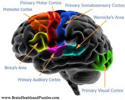
Roles of the Language in the Wernicke Area Functions and in the Intellective Functions.
A conspicuous portion of the sensorial experiences is converted in their verbal equivalent, before their storing in the memory cortex areas and before their elaboration for other intellectual purposes. In the reading process, words are not fixed in the memory as pictures, but the verbal symbols are ideal for the memorization; the information conveyed by words is converted in verbal symbols before to understand their meaning.
The sensorial area for the language interpretation in the dominant hemisphere is the Wernicke area, and this last is connected with the hearing area and with the hearing associative area of the temporal lobe. Perhaps, this union is the result of the first approach to language through the hearing. When, gradually, the visual approach to language through the reading is feasible too, the visual information is channeled in the language regions already developed in the dominant Temporal lobe.
When the Wernicke area of the dominant hemisphere is destroyed, the subject loses quite all the intellective functions associated to the language or verbal symbolism, as the reading ability, the arithmetic and logical aptitudes. Many other interpretive skills, some of which involve the Temporal lobe and the Angular Gyrus of the opposite hemisphere, are conserved. Non-dominant hemisphere may be involved in the comprehension and interpretation of music, non-verbal visual experiences (particularly of visual models), spatial relationships between the subject and his environment and, perhaps, in the interpretation of many somatic sensorial experiences associated to the arms and hands use. Thus, the dominance of the left hemisphere is related to the language—or verbal symbolism—and the opposite hemisphere can be dominant for other intellective functions.
Wernicke and Broka areas
Wernicke Area, Angular Gyrus and Broca’s Area
The association area of somatic, ears and eyes sensors inputs, which may be called “interpretive perceptive area”, are confluent in the posterior-upper portion of the temporal lobe, where the parietal and occipital lobes reach the temporal lobe. It has an important role in those superior cerebral functions that compose the intelligence. This merging cortex site of different sensorial interpretive areas is particularly developed in the dominant hemisphere—the left one in quite all the right-hand people—and it has an important role in those superior cerebral functions that compose the intelligence.
The Wernicke area is known as gnostic area, cognitive area, tertiary associative area etc. Wernicke is the neurologist that first identified the special role of this cortex portion in the intellective processes (Guyton, 1991).
The studies of the lesion in this portion of cortex reveal a preserved hearing function and the ability to distinct words, but the use of these words in a thought fails. Subjects with this lesion can read written words, but cannot decipher their meaning. An electrical stimulation of the Wernicke area in aware subjects evokes mind’s representations of high complexity. This occurs when the electrode is introduced deeply in the brain, near the thalamic connection areas. The mind’s representations that may be evoked are complex pictures memory, sometime of the childhood age; hearing hallucination, as a determinate musical partite or phrases of discourses heard previously. Therefore, it is suggested that the Wernicke area contains different mnemonic models with many sensorial involvement, or that the Wernicke area may recall memories conserved elsewhere.
The angular gyrus is located in the posterior extremity of the lateral Fissure or Silvius Fissure, behind the Wernicke area and is merged posteriorly with the visual areas of the Occipital lobe. Lesions of this area can preserve the hearing function, but the visual experiences flow from the occipital lobe to the Wernicke area is blocked. Thus, the subject can observe words and know that they are words, but he cannot understand their meaning. This condition is known as alexia or verbal blindness.
Loss of the Wernicke area produces usually a chronic dementia.
Jointly with the Temporal and the Angular Gyrus dominance, certain cortex’s portions involved in the somatic aesthesia and in controlled movements, acquire dominant function.
The premotor language area (Broca area), located in the lateral portion of the middle region of the Frontal lobe, is also dominant in the left hemisphere. It is responsible for the motor processing of the language; this area produces words throughout the simultaneous activation of the larynx, breath and mouth muscles.
Communication and Brain
There are two aspects of the communication: the sensorial, involving the hearing and the sight, and the motor, involving the vocalization and its control.
The destruction of the cortex associative areas implied in the hearing and sight functions may produce the impairment of the ability to understand written and spoken words—hearing and visual aphasia, respectively, or verbal deafness and blindness (alexia).
1. Wernicke aphasia and global aphasia. Some subjects are able to recognize both the spoken words and those written, but they are not able to interpret their meaning; this disorder is associated to an injury of the Wernicke area in the posterior portion of the upper Temporal circumvolution in the dominant hemisphere. When the lesion is spread to involve:
a. The area behind the Angular Gyrus,
b. The lower portions of the Temporal lobe, and
c. The upper edge of the Silvius Fissure, the subject suffers a quite total dementia or global aphasia.
2. The language process implies:
a. The development of the thought and the choice of the words to use;
b. The motor control of the verbal expression and the words production.
The thought development and the words choice are the main functions of the brain associative areas. Even in these activities, the Wernicke area, in the posterior portion of the upper Temporal Gyrus, plays the main role. Actually, subjects with Wernicke aphasia or with global aphasia are unable to formulate thoughts. If the lesion is less severe, the subject may preserve the capacity to formulate thoughts, but without the possibility to assemble the appropriate words. Usually, the subject collects many words arranged confusedly.
In the motor aphasia the subject is perfectly able to decide what to say, and although he can produce vocal sounds, he cannot emit words instead of sounds without meaning. This condition, known as motor aphasia, is related to a damage of the Broca’s area, located in the prefrontal cortex region and in the premotor area corresponding to the face—about the 95% of cases in the left hemisphere. From this area originate the specialized models of movement for all muscles (larynx, lips, mouth, respiratory system, etc.) that participate to the words articulation.
The final step of verbal communication is the articulation—the movements of mouth, tongue, larynx and other structures which produce words. Muscles interest in these movements are activated from the motor cortex areas corresponding to the regions of the face and larynx, while the cerebellum, the basal ganglia and the sensorial cortex participate to the muscles contraction throughout feedback mechanisms. The damage of these regions can produce a total or partial incapacity to talk distinctly.
Superior Intellective Functions of the Prefrontal Associative Area.
Before the introduction of the modern psychopharmacology for the treatment of the psychiatric disease, the prefrontal lobotomy was practiced in patients affected by severe variants of depressive psychosis—the interruption of the neuronal connections between the prefrontal areas and the other brain parts—with important improvements. The surgeon performed two breaches in the frontal sides of the skull and entered the brain with a knife blade cutting the brain in the vertical direction. Patients mind modifications were:
1. Lose of the solving problem skills;
2. Lose of the abilities to accomplish determined and longed targets throughout sequential tasks—in normal condition, if the target included motor actions, these would be actuated; otherwise the mind processes would have as result the execution of intellective analysis operations;
3. Lose of the learning abilities and of the time parallel performances;
4. Decrease of aggressiveness and belligerence; these effects were probably related to the loss of the ventral portions of the frontal lobes on the inferior faces of the brain—this portion is considered pertaining to the limbic associative cortex;
5. Disinhibiting and coarseness;
6. Speech deficits and failure to forward and preserve for long time the same thought or line of thoughts; unsettled disposition;
7. Irresoluteness and indeterminacy; the habitual actions were preserved.
The thought elaboration—or processes of highest abstraction—is another function of prefrontal areas; after prefrontal lobectomy, lab animals lost the memory abilities—perhaps because the animals were easily distracted and unable to retain the mind representations for enough time to place them in memory. The prefrontal areas capacities to stimulate the memorization—even if temporary—of many bits of information, and then to recall this information bit per bit, following the necessities of the mind processes, might explain the association of many highest cerebral functions to the intelligence expression, as
1. Previsions;
2. Future plans development;
3. Delay the sensorial signals reaction, to evaluate the information and decide appropriately the answer;
4. Consider the motor activities consequences before their execution;
5. Solve complex problem;
6. Correlate the information originating from different sources to diagnose non common diseases;
7. Control the own behavior according to the ethic.
Callosal Body and Anterior Commissure
The equivalent cortex areas of the two hemispheres, except those of anterior portions of Temporal lobes, are linked one another by the Callosal body. The cortex areas of the anterior portions of Temporal lobes— including the Amygdala—are linked one another by fiber traversing the anterior Commissure.
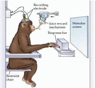
Recently, specific researches led with the support of psychological methods, have demonstrated the importance of the Callosal body and anterior Commissure functions. These functions are showed by the description of one of these researches. A monkey undergone to a Callosal body and Optic Chiasm division; next the animal was taught to recognize different types of objects with the right eye, keeping the other closed. A battery of tests is administered to the animal to establish if it is able to recognize the same objects with the left eye, keeping the other closed. The results show that the animal fails to recognize the same objects with the left eye, but if the experiment is reproduced on another monkey, leaving the Callosal body intact and dividing only the optic Chiasm, the identification of the objects by a hemisphere affects the identification of the same object by the other hemisphere, invariably. Therefore, one of the Callosal body and anterior commissure function is to allow the transition of information from a hemisphere area to the correspondent other.
1. The Callosal body division prevents the information transmission from the Wernicke area of the dominant hemisphere to the motor cortex of the contralateral hemisphere. So, the intellective functions of the brain, mainly localized in the dominant hemisphere (more often the left hemisphere), can neither control the right motor cortex, nor the voluntary motor activity of the left hand and arm, even if the subconscious movements of the different segments of this limb are normal.
2. The Callosal body division prevents the visual and the somatic aesthesia information transmission from the right hemisphere to the Wernicke area of the dominant hemisphere. So, the information of these sensorial modalities that originate from the left side of the body cannot reach the Wernicke area, and therefore they cannot be used in the cerebration processes.
3. In an adolescent to whom the Callosal body had been completely cut, leaving the anterior commissure intact was observed that the aware brain resulted split in two separate halves. Only the left half could understand the spoken words, being the dominant hemisphere. The right hemisphere could understand the written words and react with a motor answer, without revealing the reason to the left hemisphere. The effect was different when an emotional answer had been evoked in the right half of the brain, because an emotional answer was evoked in the left hemisphere likewise. It was related to the preserved communication of the Temporal lobes and other areas of the lower regions of the brain between the two hemispheres throughout the anterior Commissure. For instance, when the written command “kiss” was showed to the right hemisphere of the patient, he answered “no”. This answer required the Wernicke and language motor areas of the left hemisphere. Nevertheless, when he was asked why, he did not know. The two hemispheres have independent capacities of consciousness, memory, communication, and of motor activity control. The Callosal body function is necessary for the cooperation of the hemispheres, and the anterior Commissure is important to unify their emotional answers.
Thoughts, consciousness, and memory
Certainly each thought requires the simultaneous activation of many cortex cerebral areas, of the thalamus, limbic system, and of the brainstem reticular formation. Perhaps, some types of mind activities are related to the subcortical centers. Paradigmatic is the pain experience, since the stimulation of the cortex rarely produces a modicum pain, while the stimulation of certain areas of the hypothalamus and mesencephalon seems to produce a strong pain. Certain types of thoughts require first the cortex function and they involve the visual representations, since the loss of the visual cortex determines the total incapacity to perceive visual shapes or colors.
Thus, a definition of thought considering the nervous activity might be the following: a thought is the product of the simultaneous activation of many portion of the nervous system, of the thalamus, limbic system, and of the rostral portion of the brainstem reticular formation, in a determined sequence. This is the holistic theory of the thought. The activation of the limbic system, thalamus, and of the reticular formation, produces the general nature of the thought, providing it of painful or pleasant characters, raw experiences of the different sensorial types with different body localization, etc.
The cortex activity produces probably the detailed character of the thought, as the specific localization of senses in different body parts, and of objects in the visual field, and defined models of sensations as objects geometric shapes or tissues’ frames, which compose a temporal global cognitive image.
The consciousness may be defined as the bags of data immediately available for the function in the environment and for the abstractions; conserved, renewable or modified in relation to the importance invested in a particular state of life, present or with temporal projection; paralleled to the sensorial composition (reference to pleasant & painful characters), and distinct from the memory while using its contents.
Hippocampus
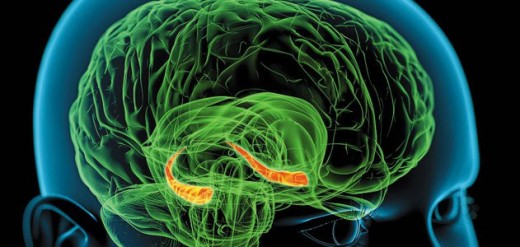
Hippocampus and memory
The Hippocampus is the most medial portion of the Temporal cortex where this encircles the inferior-medial edge of the hemisphere and goes up to the inferior surface of the lateral ventricle. The hippocampus is one of the most frequent neurosurgeons’ targets in the epilepsy treatment. The hippocampi removal does not affect the memory of the subject concerning the information memorized before the surgery. Nevertheless, after the hippocampi removal subjects reveal difficult to transfer verbal and symbolic types of immediate memories in long-term memories lasting few minutes. Therefore, these subjects are unable to have lasting memories of that information that are the base of the intelligence. This condition is called anterograde amnesia. The hippocampus importance in new memory development is related to its anatomical location, as it is the main yielding pathway of the gratifying and punishing areas of the limbic system. These are located in different structure of the brain’s basal regions and transmit information to the hippocampi. Those sensorial experiences or mind representations that evoke pain or aversion stimulate the punishing centers, while those pleasant, glad or satisfying experiences stimulate the gratifying centers.
From these complex processes depend the mood and the subject’s motivations or mind representations that are pleasant or unpleasant. The hippocampus, particularly, and the medial dorsal nuclei of the thalamus, another structure of the limbic system, are important in the decisional process on which experience or other psychic activities, in terms of gratification or punishing, are important enough to be memorized. Likewise, lesions in other portions of the Temporal lobe other than the hippocampus are followed by a reduced capacity to memorize new information. This can be related to two factors:
1. Possibility that they are portions of the Temporal lobe associated with the hippocampus function, so their lesion might compromise the normal memory consolidation process, and
2. The Wernicke area might be damaged—located in the Temporal lobe and site of the main intellective activity. Lesions of this area might reduce the capacity to memorize new information as the memory consolidation requires an analysis of this information that allows the association with the similar one.
Retrograde amnesia
Retrograde amnesia indicates the inability to evoke old information—as from the lasting memories—even if the subject is aware that they are still in the memory. When a subject is affected by amnesia, the loss of recent information or events is major than that of old or remote events. Perhaps this occurs because the old memories have been represented so many times that some their elements are deposited in large portions of the brain. Some subjects with hippocampus lesions and anterograde amnesia present some degrees of retrograde amnesia too. This suggests that these two kinds of amnesia are correlated, at least partially, and they can be both produced by hippocampus lesions. Several cases where some thalamic lesions produced retrograde amnesia without association with the anterograde amnesia were described. This might be explained by the thalamus main role in looking for information placed in memory, allowing the subject to extract memories. The process of memorization requires the placing step and the capacity to search and find the information in next steps.
Hippocampus disconnection from some types of learning
Subjects with hippocampus lesions or other portions of the Temporal lobe do not have difficulties for leaning body’s activities that require attention without involve verbal processes or other symbolic types of intelligence. For instance, these subjects are still able to learn complex physical works, as those of some jobs, and sports activities that require special abilities. In these cases the learning process is related to the repetition of the body activities rather than abstractions reiteration.
Limbic System
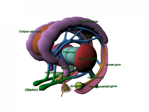
Limbic system
The term “limbic” means “to the edge, to the border, to the boundary” and was first used to name some structures that surround the basal regions of the brain. With the knowledge advance of the limbic system, the meaning of this name has been extended to indicate the entire complex of neuronal circuits that control the motivational pushes and the emotive behavior of the subject. A relevant portion of the limbic system is constituted by the hypothalamus and by the correlated structures. Other than their roles in the behavior, these areas control many interior conditions of the organism, as the body temperature, the body liquids osmolality, the body weight, the starving and the thirst, etc. These are called as vegetative functions and their control is strictly related with the behavior.
Functional anatomy of the limbic system and relation with the hypothalamus
The limbic system is constituted by a complex of structures strictly connected one another, and located on the brain basal areas. In the middle of these structures is placed the hypothalamus, which is considered as a distinct formation from the limbic system. Nevertheless, under a physiological point of view, it is one of the central elements of the system. The hypothalamus is surrounded by the subcortical structures of the system, including the septum, the paraolfactory area, the epithalamus, and the anterior nuclei of the thalamus, part of the basal ganglia, hippocampus and the amygdala. The limbic cortex is around the subcortical limbic structures and is constituted by a cerebral cortex ring that 1) originates in the eyes frontal area on the ventral surface of the Frontal lobes, 2) continues upon in the subcallosal gyrus, below the anterior margin of the callosal body, 3) continues in the cingulate gyrus above the callosal body on the medial face of the hemisphere, and at the end 4) goes posteriorly to the callosal body and down on the ventromedial surface of the Temporal lobe, and continues in the parahippocampal gyrus to terminate with the uncus. So, on the medial and ventral surfaces of each hemisphere there is a ring of cortex, mostly of paleocortex that encircles a group of deep structures strictly connected with the global behavior and with the emotions. This ring of cortex acts as a pathway for the bidirectional communication and association between the neocortex and the different limbic subcortical structures. It is important to stress that many of the behavioral functions that originate in the hypothalamus and from other limbic structures occur through the mediation of the reticular nuclei and their associated nuclei of the brainstem. The stimulation of the excitatory portion of the reticular formation exalts the somatic and cortical excitability degree, and the greatest part of the hypothalamic signals for the autonomic nervous system control, is transmitted by nuclei located in the brainstem. An important communication pathway between the limbic system and the brainstem is the prosencephalic medial fasciculus, which from the septum region and the eyes frontal cortex reaches down the hypothalamus and the reticular formation. It is a fasciculus made by fibers of both the directions, which constitutes a major communication system. A second communication via is made of short paths between the reticular formation, the thalamus, the hypothalamus and the great part of the other contiguous areas of the basal brain.
Hipothalamus
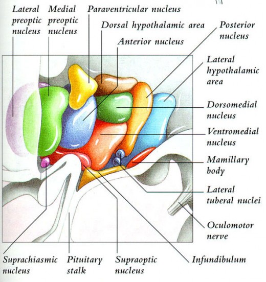
Hypothalamus, main output channel of the limbic system
The hypothalamus communicates with all the levels of the limbic system. The hypothalamus and structures associated to him send signals in three directions:
1. Down in the brainstem, mainly at the reticular formation of the midbrain, pons and medulla oblongata;
2. Up toward many highest areas of the diencephalon and telencephalon, mostly at the anterior thalamus and to the limbic cortex; and
3. At the infundibulum to control the secretory activity of both the anterior and posterior hypophysis.
Therefore, the hypothalamus, that represents less than the 1% of the brain, is one of the most important output channels of the limbic system. It controls the greater part of the endocrine activities, of the vegetative functions of the body and many emotional behaving aspects too.
Behavioral function of the hypothalamus and of the limbic structures associated
The stimulation or the lesions of the hypothalamic structures usually have deep consequences on the emotional behavior both in the animals and in the man. In animals, some effects of the behavioral stimulation are:
1. The stimulation of the lateral hypothalamus produces not only hunger and thirst, but it induces an increase of the general levels of animals’ activity too, sometimes reaching a clear behavior of angry and fights.
2. The stimulation of ventromedial nuclei and of the surrounding areas produces effects opposed to those induced by the stimulation of the lateral hypothalamus—that is, a satiety feeling, decreasing of feeding, and quiet state;
3. The stimulation of a slight area of the periventricular nuclei, immediately adjacent to third ventricle (or the mesencephalon central area that continuous with this hypothalamic portion too), generates behavioral reactions of fearing and punishing;
4. The sexual impulse can be evoked by the stimulation of different hypothalamic areas, especially those located on the anterior and posterior extremes of the hypothalamus.
Lesions of the hypothalamus generates opposite effects to those above reported. For instance:
1. Bilateral lesions of the lateral hypothalamus reduce to zero the feeding and drinking, often to the inanition state. Nevertheless these lesions cause an extreme apathy of the animal, with loss of many of its activity manifestations;
2. Bilateral lesions of the ventromedial hypothalamic area generate opposite effects to those presented above: excessive feeding and drinking, hyperactivity and often a continuous state of aggressiveness, with frequent angry outburst for the least provocation.
Likewise, lesions or stimulations of other regions of the limbic system—especially of the amygdala or of the septum areas—and of mesencephalon areas generate similar effects to those showed above for the hypothalamus.
Limbic roles in the gratifying and punishing
The hypothalamus and other limbic structures are particularly involved in varying the affective tone and in the sensorial experiences—or, in the pleasant and unpleasant feelings that accompany them. These affective tonalities are called gratifying (or recompense) feelings and punishing feelings. Other nomenclatures can be used as, of satisfaction and aversion, respectively. While the electrical stimulation of some regions of the limbic system produces in the animal pleasure or satisfaction, the stimulation of other areas evokes terror, pain, fear, defensive reactions and escape, etc.
Gratifying centers
The technique used for the localization in the brain of the specific areas for the gratification and the punishing use a monkey in a container with a handle that controls with a downward movement the activation of an electrical stimulator. The electrodes are applied in sequential steps in different areas of the encephalon that the animal can stimulate with the handle movements. If the stimulation of a particular area evokes a gratifying feeling, then the animal will push down the handle repetitively, sometimes even thousands times in one hour. When the animal is in the condition to choose between the handle stimulation and the feeding, the animal will continue to stimulate itself. With this method the main gratifying brain centers was located along the prosencephalic medial fasciculus, especially in the lateral and ventromedial nuclei of the hypothalamus. It may appear strange that the lateral nuclei are between the gratifying—actually they are the most potent--but stimulating them strongly, they evoke anger.
Generally, a light stimulation evokes a gratifying feeling, while strong stimulations evoke a punishing feeling.
Less powerful gratifying centers, maybe secondary, are in the septum, amygdala, in certain thalamic and basal ganglia areas, and lower in the basal portion of the mesencephalon tegmental.
Punishing centers
The handle can have the reverse function with the circuit interruption with the downward movement. In this case, the animal does not use the handle, and in this way it does not discontinue the stimulation, when the electrode is in a gratification area, but when it is in a punishing area, the animal learns to stop the stimulation. The activation of these areas evokes all the signs of unpleasant, disquieted, terror, and punishing feelings. If it is protracted for 24 hours or more it may produce in the animal a grave state of suffering that can produce the death.
With this technique the main punishing centers and those of the escape impulses were located in the periaqueductal center area of the mesencephalon and they extend up in the periventricular areas of the hypothalamus and of the thalamus. Punishing centers are less potent and they were localized in the amygdala and in the hippocampus.
It is interesting that the stimulation of the punishing centers usually inhibits completely the gratifying centers and of the pleasure, showing that the punishing feeling and of fear can have the priority on those of gratification and pleasure.
Gratifying and punishing importance in the behavior
Fairly all the actions of the men are in some way related to the gratifying or punishing feelings. If something gratifying were performed, the tendency would be to continue doing it; instead if something punishing were operating, it would be ceased. Therefore, the gratifying and punishing centers are the most important systems of the human activities and pulsing regulation.
Angry and tranquility
Angry
The angry is a model of emotional behavior that involves the hypothalamus and many other limbic structures and presents well defined characters. The angry can be evoked by a strong stimulation of the encephalic centers of the punishment, especially in the periventricular area of the hypothalamus or of the lateral hypothalamus, and it is characterized by 1) defensive posture, 2) extension of the claws, 3) tie raises, 4) sibilates, 5) salivation, 6) to growl, and 7) hair erection, wide open eyes and pupils dilated. Even to the minimal provocation the animal reacts with a ready and ferocious attack. The stimulation of the most rostral areas of the punishing centers—in the preoptic areas on the middle line—produces mostly fear and anxiety, associated to escape tendency. In the normal animal the angry are repressed mainly by the antagonistic activity of the ventromedial nuclei of the hypothalamus. However, to hold it in an inhibited state contributes the hippocampus, the amygdala and the anterior portions of the limbic cortex, mainly of the subcallosal gyrus and of the anterior portions of the cingulate gyrus. If these portions of the limbic system are damaged or destroyed, the animal (or the man) becomes very susceptible to the angry accesses.
Tranquility and compliance
The stimulation of the gratification centers evokes models of emotional behavior opposed to the precedent, as the tranquility and the compliance.
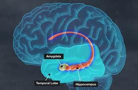
Amygdala
The amygdala is a complex of nuclei located immediately beneath the medial surface of the cerebral cortex in the anterior pole of each Temporal lobe. It has many bidirectional connections with the hypothalamus. In many animals this complex is involved in a large part of the associative processes between the smell and other sensorial types. One of the principal branches of the olfactory “benderella” conduces directly to the corticomedial nuclei, a portion of the amygdala placed under the cortex in the piriform area of the Temporal lobe. But in the men another portion of the amygdala has reached a very high degree of development in the olfactory area: it is the portion of the basal lateral nuclei that have important roles in many behavioral activities not associated generally with olfactory stimuli. The amygdala receives signals from all the limbic cortex areas and from the neocortex of the Temporal, Parietal and Occipital lobes, especially from the associative areas of hearing and sight. For these multiple connections, the amygdala was defined the window through which the limbic system sees the person in his relation with the environment. The amygdala transmits its signals 1) to these cortex areas, 2) to the hippocampus, 3) to the septum, 4) to the thalamus, and 5) especially to the hypothalamus.
Effects of the amygdala stimulation
In general, the stimulation of the amygdala can produce almost all the effects that follow the hypothalamic stimulation, and many other. The effects that are mediated by the hypothalamus comprise: 1) increase or decrease of the blood pressure, 2) increase or decrease of the cardiac pressure, 3) increase or decrease of the motility and of the gastroenteric apparatus secretory activity 4) defecation and urination, 5) pupils dilatation or, rarely, constriction, 6) hair erection, 7) hormones secretion of the anterior hypophysis between which, mainly, the gonadotropins and the ACTH.
Other than these effects mediated by the hypothalamus, the amygdala stimulation can evoke different types of involuntary movements: 1) tonic movements, as the head extension and the body curving; 2) circular movements (manipulating); 3) clonic movements, rhythmic, and 4) different types of movements associated with the act of smelling or with the assumption of foods, as the lick up, mastication movements and deglutition.
Further, the stimulation of certain nuclei of the amygdala can determine, rarely, angry, fear, escape reaction, and other signs of the punishing feeling, with a picture similar to that of the hypothalamic starting angry. The stimulation of other amygdala’s nuclei can evoke, instead, reaction of gratification and pleasure. In conclusion, the activation of other parts of the amygdala can have effects on the sexual function and reproductive, as the penis erection, copulation movements, ejaculation, ovulation, uterine contraction and premature delivery.
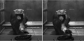
Effects of the bilateral ablation of the amygdala—the Klüver-Bucy syndrome
If in a monkey the anterior portion of both Temporal lobes, other than the corresponding regions of the temporal cortex are destroyed, the amygdales, located deeper in these lobes are destroyed too. A complex modification of the behavior, under the name of Klüver-Bucy syndrome is the result, and it comprises: 1) excessive tendency to the oral exploration of objects, 2) absence of fear, 3) decrease of aggressiveness, 4) compliance, 5) feeding habits changes, often an herbivore become carnivore, 6) psychological blindness, 7) often, excessive sexual pulse. The characteristic picture is that of an animal that does not have fear of nothing, shows an extreme curiosity for everything, forgets quickly, puts everything in its mouth, and often it has a sexual impulse so strong that it is pushed to couple with sexual immature animals and with animals of other species too. Men present a syndrome similar to that observed in the monkey.
Functional role of the amygdala
The amygdala seems to have the function of a control center of the behavior, and operates at a semi-unconscious level. It seems that it projects to the limbic system the current state of the subject in relation to the environment and to his psychic dimension. On the base of this information the amygdala would contribute to the determination of the behavioral model of the subject answer, so that this would be adequate to the contingencies.

Limbic cortex function
Perhaps, the less known portion of all the limbic system is the limbic cortex, or the ring of cerebral cortex edging the subcortical limbic structures. This cortex part works as a transition area through which signals are transmitted to the limbic system from other cortical regions. Therefore, the limbic cortex has the function of associative areas for the behavior control. The stimulation of different parts of the limbic cortex did not produce enough elements for an acceptable hypothesis on their function. As for many other portions of the limbic system, even for the limbic cortex the stimulation of its different portions can evoke all the behavioral models mentioned above. Concerning the ablation experiments, the limbic cortical areas whose ablation can cause persistent modification of the behavior in the animal are few. In many cases the ablation has only limited effects. Between the areas whose ablation showed specific effects are the following:
1. Temporal cortex ablation. When the anterior Temporal cortex is removed bilaterally, even the amygdala is invariably compromised. The Klüver-Bucy syndrome appears then. The animal assumes a consuming behavior, it explores the environment and every object, it is motivated from strong sexual impulses even with not ideal animals and with inanimate objects, and it loses the fear—it is at the same time compliant.
2. Posterior frontal orbital cortex ablation. The bilateral ablation of the posterior frontal orbital cortex, usually produces in the animal insomnia and motor agitation, and makes it unable to stay seated pushing it to move around continuously.
3. Subcallosal gyrus and anterior portion of the cingulate gyrus ablation. The subcallosal gyrus and the anterior portion of the cingulate gyrus are the limbic cortex portions that put in conjunction the prefrontal cortex and the subcortical limbic structure. Their bilateral ablation releases from every prefrontal cortex inhibitory influence, the centers of the angry in the septum and in the hypothalamus. Thus, the animal becomes aggressive and much more susceptible to angry accesses.

")


