What is the Urinary System


Urinary system: This are made up of organs that produces and excretes urine from the body. The organs of the urinary system includes a kidney, a pair of ureter, urinary bladder and a urethra.
THE KIDNEY: The kidney is a reddish- brown, bean shaped organ having a smooth surface and are enclosed in a tough, fibrous capsule. The kidney lies on the either side of the vertebra column in a depression high on the posterior wall of the abdominal cavity. The kidney are placed retroperitoneally, meaning they are behind the parietal peritoneum and against the deep muscle of the back. The kidney are held in position by the connective tissue and the masses of adipose tissue, which surrounds them. The left kidney is higher than the right kidney.

STRUCTURE OF THE KIDNEY: The kidney comprises of two distinct region known as the renal medulla (inner) and a renal cortex (outer). The renal medulla is composed of conical masses of tissue called renal pyramids, which appears striated, while the renal cortex forms a shell round the medulla and dips into the medulla between renal pyramids, thereby forming a renal column. The lateral surface of the kidney is convex while its medial side is concave; the medial side leads into a hollow chamber known as the renal sinus. The entrance through the renal sinus is called the hilum through which blood vessels, lymphatic vessels and ureter passes into and leaves the kidney. The superior end of the ureter expands to form a funnel shaped sac called the renal pelvis right inside the renal sinus. The pelvis sub divides into three tubes known as major calyces, which also subdivides into other several minor calyces. Renal papillae projects into the renal sinus from its wall.
The nephron is the functional unit of a kidney, which is about one million in a kidney. It consists a renal corpuscle and a renal tubule. The renal corpuscle consist a glomerulus and a glomerular capsule, while the renal tubule is a passage through which fluid flows on its way out of the body. It leads away from the glomerular capsule and becomes highly coiled called the proximal convoluted tubule. The glomerular capsule receives a filtered fluid from the glomerulus
FUNCTIONS OF THE KIDNEY
1) The kidney maintains homeostasis by removing metabolic wastes from the blood and excreting them.
2) The kidney help regulate red blood cell production, blood volume and blood pressure, composition and pH of the body fluid.
URETER: each of the ureter is a tube, which begins as the funnel shaped renal pelvis. The ureter extends downward behind the parietal peritoneum and runs parallel to the vertebral column. The ureter course forward and medially joining the urinary bladder from underneath. The ureter is made up of three main layers.
(i) The inner layer (mucous coat) is continues with the linings of the renal tubules and the urinary bladder.
ii) The middle layer (muscular coat) consist a smooth muscle fibers.
iii) The outer layer (fibrous coat) is a connective tissue.
The muscular wall of the ureter propels the urine. The muscular peristaltic wave that originates in the renal pelvis forces urine along the length of the ureter. When the peristaltic wave reaches the urinary bladder, a jet of urine spurts into the bladder. A fold of mucous membrane act a valve, it allows urine enter the bladder but prevents it from going back up.
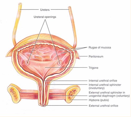
THE URINARY BLADDER: the urinary bladder is a hollow and muscular organ that stores urine and forces it into the urethra. It is located within the pelvic cavity, behind the symphysis pubis and beneath the parietal peritoneum. The wall of the urinary bladder are made up of four layers; the mucous coat, the sub-mucous coat, the muscular coat and the serous coat. The urinary bladder holds as 600 milliliters of urine before stimulating pain receptors, but the urge to urinate usually begins when it contains about 150 milliliters. When the bladder empties, its wall forms a fold and it becomes smoother as urine fills it. At the internal floor of the bladder is a triangular area known as the trigone, this trigone has an opening at each of its three angles. There are ureter openings at the posterior base of the trigone, and at the anterior apex of the trigone called the neck of the bladder. It contains the opening into the urethra. The neck of the bladder forms an internal urethral sphincter, and when the muscle around the urethral sphincter contracts it prevents the bladder from emptying until pressure within the bladder increases to a certain level.
THE URETHRA: This tube carries urine from the urinary bladder to the outside. The wall of the urethra is lined with a mucous membrane, a thick layer of smooth muscle tissue and numerous mucous glands called the urethral glands, which secretes mucus into the urethra canal.
FORMATION OF URINE
The formation of urine takes place in the nephron. It begins with the filtration of plasma by the glomerular capillaries, a process called glomerular filtration. Glomerular filtration produces about 180 liters of fluid more than four times the total body water every 24 hours. Two other process contribute to urine formation. They are the tubular reabsorption, which moves substances from the fluid back into the blood with the peritubular capillary, and a tubular secretion, which moves substances from the blood within the peritubular capillary into the renal tubule. In the tubular reabsorption, the kidney selectively reclaims the right amount to substances such as water, electrolyte, and glucose that the body requires while waste and excess substances leaves the body. In tubular secretion, some substances, which the body must eliminate such as hydrogen ion and certain toxins, are removed faster than through filtration alone. The final product of these three processes, glomerular filtration, tubular reabsorption, and tubular secretion is known as urine.
THE ELEMINATION OF URINE
After urine is formed in the nephron, it passes from the collecting duct through openings in the renal papillae and enters the calyces of the kidney, then passes through the renal pelvis and the ureter carries it to the urinary bladder, which takes it to the urethra, and then the urethra takes it to the outside.
Related

Instrumental Methods of Examination, X-rays and Radioisotopic diagnostics in Urological diseases

Symptoms of Kidney Disease in Cats

Pyelonephritis - E. Coli Kidney Infection

Does Your Dog Have Bladder Stones? An Interview With a Veterinarian

Urine Colors Chart : Medications and Food Can Change Urine Color


