Let's tackle glomerular Nephritis in full

Glomerulonephritis
the soul talkers

Biblical love psychology

Acute Glomerular Nephritis
Aetiology. Acute diffuse glomerulonephritis usualy develops acute infectious diseases, such as tonsillitis, scarlet fever, acute respiidiseases, pneumonia, and otitis.'Especially imp7)TtanTare diseases c by group A haemolytic streptococcus, most frequently of type XInephritis can arise also after infectious diseases caused by other bat e.g. pneumococci or staphylococci. Acute nephritis sometimes de1following overcooling, especially in damp weather. Cases were repor acute nephritis developing after vaccination.
Pathogenesis. Acute nephritis typically arises not during an infe disease but only following a period of time, usually 2-3 weeks latejtempts to isolate the streptococcus from the kidney tissue end in fi Thus, the onset of acute nephritis usually coincides with the periodantibodies to streptococcus are produced. This indicates that nephritis is not simply an infectious disease but an infectious a disease.
It is suggested that bacterial antigens, that get into the blood dur fection, injure the kidney tissue, whose affected proteins act as an a to stimulate the production of the corresponding antibodies I reticuloendothelial system. The antigen-antibody complexes are fi the endothelial and epithelial cells of the renal glomeruli and also ' basal membrane of the glomerular capillaries to cause their injury kidneys are always involved in acute diffuse glomerulonephritis a glomeruli are equally affected. This distinguishes the affection fron nephritis and confirms its allergic nature.
It is necessary to note that both the glomerular capillaries and ves / the other organs and tissues are affected in acute glomerulonepNephritis is thus the general vascular disease. Cases have been des when in the presence of a marked clinical picture of the disease (oehypertension), the urinary symptoms were insignificant or absent. B rule, the glomerular apparatus of the kidneys is affected in acute ne which is explained by the specific character of their function as t cretory organ.
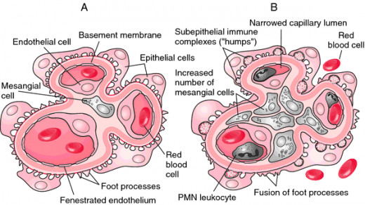
Pathological anatomy. The kidneys of those who died from acute nephritis are of size or slightly enlarged; their colour is brown or greyish-brown. Malpighi corpuscle form of small tubercles, can be seen on section. Microscopy of the renal tissue in th stage of the disease reveals enlarged and hyperaemic glomeruli; during further progrei disease, microscopy reveals ischaemia of the glomeruli due to spasm of the capillar fibrinoid swelling of the capillary walls, proliferation of their endothelium, accumul coagulated proteinous exudate in the space between the capillary loops and the glo capsule, blood stasis, thrombosis of the capillary loops, and haemorrhages. Path changes occur in both kidneys in all cases. Epithelium of the renal tubules is affe larkedly. Intra- and extracapillary glomerulonephritis is mostly differentiated; depending on le character of the inflammation, glomerulonephritis may be exudative (serous, fibrinous, nd haemorrhagic) and productive.
At later stages of the disease, inflammatory phenomena subside in renal tissue, prolifera-lon of endothelium in glomerular loops decreases, and patency of the capillaries is restored.
Clinical picture. The clinical picture of acute glomerulonephritis is mite specific and is determined by the main three syndromes: oedema, irterial hypertension, and changes in the urine (haematuria and prolinuria). The patients would usually complain of oedema, which arise first an the face, under the eyes, and then extend onto the entire body and the extremities. Development of oedema is explained by disordered capillary permeability and aldosterone hypersecretion by the adrenal cortex, Headache and heaviness in the head are frequent symptoms. They are explained by increased arterial pressure and, in some cases, intracranial pressure. Vision can be deranged due to spasm of the retinal vessels and haemorrhages into the retina. Many patients complain of general fatigue and reduced work capacity.
In the presence of a pronounced oedema and massive pleural effusion, and when the heart muscle is overloaded due to markedly increased arterial pressure, patients with acute nephritis suffer from severe dyspnoea,sometimes with attacks of asphyxia (like in cardiac asthma).
The patient with acute nephritis would often complain of dull lumbar pain. The gravity of the disease depends on the degree of oliguria. The diuresis decreases while the patient may have frequent tenesmus. Complete anuria occurs in some cases. If haematuria is marked, the urine looks like meat wastes.
Inspection of the patient reveals his specific appearance: pallid skin, oedematous face, swollen eyelids, and oedema of the trunk. Some patients assume the forced semireclining or sitting position because of pronounced dyspnoea. Renal eclampsia occurs in grave cases. The onset of an;eclarrtp-sia attack is heralded by increasing arterial pressure and a severe headache. The extent and the character of oedema can be established by palpation. The pulse of the patient should also be felt. Acute nephritis is characterized by a tense pulse which is sometimes slow. The apex beat is somewhat shifted to the left and increased due to myocardial hypertrophy which soon develops in the presence of arterial hypertension.
Percussion of the chest in the presence of generalized oedema reveals free fluid in the pleural cavity (transudate) and congestion in the lung root region (dulled tympany). The left border of the heart extends beyond the corresponding midclavicular line.
Normal or harsh respiration is heard by auscultation. In the presence of pronounced congestion, dry and moist congestive rales are heard.
Auscultation of the heart reveals bradycardia (due to the reflex transmitted in increased pressure from the aorta onto the vagus nerve through n. depressor).
The first sound is sometimes decreased at the heart apex. If the heart muscle is much overloaded, the gallop rhythm is heard. The second sound is usually accentuated over the aorta due to increased arterial pressure.
X-ray studies of the chest confirm the presence of pleural effusion and congestion in the lung roots. Dilatation and hypertrophy of the left ventri cle are clearly determined (the heart apex is rounded). Sphygmomanome-try is of great help in establishing a diagnosis. It reveals one of the main symptoms of acute nephritis, i.e. arterial hypertension. Systoiic pressure increases to 200—220 mm Hg, but in some cases it is not so high. Diastolic pressure increases to 100-160 mm Hg almost in all cases.
Electrocardiography reveals signs of hypertrophy and overload of the left-ventricular myocardium. The amplitude of ECG waves decreases in pronounced oedema of the trunk.
Changes in the urine are characteristic of acute nephritis. During development of oedema, diuresis usually decreases to oliguria. The urine of patients with acute nephritis usually contains much protein and erythrocytes due to the increased permeability of the renal capillaries. Ifhaematuria is pronounced, urine can be reddish-brown (the colour of meat wastes). Microscopy of the urinary sediment usually reveals the presence of casts (mainly hyaline casts) and cells of renal epithelium. The nitrogen excretory function of the kidneys is usually not affected in acute nephritis. Nitrogenous slags can only accumulate in the blood in serious cases attend ed by anuria. The clearance tests reveal more or less considerable reduction of glomerular filtration.
The infectious allergic character of acute glomerulonephritis is confirmed by immunological shifts: the content of a2- and 7-globulins in the blood increases during the acute period.
Acute glomerulonephritis often proceeds without pronounced symptoms which make it difficult to identify it and hence to prescribe the ap propriate treatment. But mild and indistinct forms of glomerulonephritis, like acute forms of this disease with classical clinical symptoms, give rise to chronic glomerulonephritis, unless the appropriate therapy is given.
The gravest and even dramatic (Tareev) complication of acute
glomerulonephritis is renal eclampsia which occurs in 4—10 per cent of patients (mostly in children and women). During a convulsive attack, the patient may be heavily contused or his ribs may be fractured. Cases were reported where patients died from cerebral circulatory disorders or lung oedema; true, such cases are rare. Attacks of eclampsia usually leave no consequence. It is interesting to note that eclampsia sometimes serves as a stimulus to a rapid regress of the disease and patient's recovery.
Course. Acute glomerulonephritis usually lasts only a few weeks or months. The first sign of beginning recovery is resolution of oedema and : further decrease in arterial pressure. Small haematuria and proteinuria can persist for months following disappearance of the main symptoms. Some patients do not recover completely and the disease becomes chronic.
Treatment. Patients with acute nephritis should be taken to hospital. It is important that the air in the ward should be warm and dry; drafts should be absent. Sodium chloride intake should be restricted to 0.5-1,5 g a day, which promotes resolution of oedema and normalization of arterial pressure. Protein intake should also be slightly decreased (at the expense of meat protein).
Prednisolone and other corticosteroid hormones having anti-allergic and anti-inflammatory properties are efficacious means of pathogenetic therapy of acute nephritis. Rauvolfia is given to control hypertension; furocemid and other diuretics should be given to remove oedema.
Prophylaxis. Hardening of the body and also thorough sanation of the infection foci (carious teeth, chronic tonsillitis, sinusitis, and the like) are required.
Chronic Glomerulanephritis
Aetiology and pathogenesis. Chronic diffuse glomerulonephritis is a relatively common disease. It is often secondary to the acute form of this disease if the patient is not timely and properly treated. In other patients, chronic glomerulonephritis occurs suddenly, without acute nephritis in their anamnesis, but it can be suspected that the chronic disease was preceded by acute nephritis which however was latent, without manifest symptoms, and therefore not identified in proper time.
Chronic diffuse glomerulonephritis can sometimes be secondary to nephropathy of pregnancy which was not treated properly.
Great importance is now attached to the auto-immune mechanism in the pathogenesis of chronic glomerulonephritis. Antibodies to altered proteins of the renal tissue are probably formed in patients with the disease, in addition to formation of antibodies to streptococcus. This maintains the inflammatory process in the kidneys and is the cause of chronic progressive course of the disease.
Pathological anatomy. The kidneys are not enlarged, or enlarged only slightly during the first period of the disease, which lasts several years. In the final stage of the disease, the kidneys are markedly diminished in size, their surface is granular, the renal tissue is firm (arteriosclerotic kidney). Microscopy in chronic nephritis reveals mostly intracapillary inflammation in the glomeruli with gradual obliteration of the capillary loops and the capsule cavity and conversion of the glomerulus into a scar or hyaline node.Dystrophic changes occur in the epithelium of the renal tubules.
Clinical picture. Two periods can be easily distinguished in the of the disease: the first period, when the nitrogen secretory function of the kidneys is impaired only insignificantly (the stage of renal compensation), and the second period, during which this function is affected substantially (the stage of renal decompensation).
The symptoms of the first period are the same as in acute nephritis. The patient may complain of weakness, more or less persistent headache, dizziness, and oedema. But the gravity of these symptoms is usually less significant than in acute nephritis. The disease is often asymptomatic and is only revealed accidentally, during out-patient examination. Objective studies help to establish increased arterial pressure and hypertrophy of the left ventricle. Urinalysis reveals proteinuria and cylindruria. The presence of waxy casts is especially important diagnostically. The urinary sediment usually contains a small quantity (less frequently, considerable quantity) of leached erythrocytes. The blood serum cholesterol content is increased. More or less significant hypoproteinaemia is observed due to permanent teinuria.
Symptoms of the second, or final, period of the disease gradually due to the progressive nephrosclerosis. Low indices of clearance tests indicate decreased quantity of functioning kidney tissue. The filtration capacity of the kidneys remains unchanged for a long time and only decreases during exacerbation of the process. The concentration capacity of the kidneys gradually decreases along with the decrease in the specific gravity of the urine. Removal on nitrogenous slags from the body is maintained during this period by polyuria (evacuation of much liquid from the body). Nocturnal diuresis increases by the compensatory mechanism as well: it is two thirds-one half of the daily diuresis (nycturia). As the concentration capacity of the kidneys isaffected to a greater extent, the specific gravity of the urine becomes lowand its variations between 1.009 and 1.011 during the course of the day(and under the effect of dry food) are insignificant (isohyposthenuria). The content of nitrogenous slags in the blood of patients (urea,creatinine, indican) increases during this period.
Symptoms of uraemia develop: weakness becomes more considerable, the patient complains of lassitude, headache, nausea, skin itching, unpleasant ammonium breath, and impaired vision. Not long before death, the patients develop uraemic coma.
Course. Chronic nephritis usually lasts from 2-3 to 10-15 years. The first period of the disease (renal compensation) is long; the second period (decompensation) is shorter. During the course of the disease, there occur more or less prolonged periods of exacerbation, which are usually provoked by cooling or infections; exacerbations are followed by remissions.
Several clinical forms of chronic glomerulonephritis are differentiated by the character of its course and prevailing symptoms. Thenephrotic form is characterized by oedema, the urinary syndrome, and a comparatively rapid course. The hypertensive form is comparatively benign and is characterized by the hypertensive syndrome and insignificant changes in the urine. The mixed form is characterized by oedema, changes in the urine, and arterial hypertension. This form of glomerulonephritis is the gravest and comparatively rapid: a pronounced renal failure develops in 2-3 years. Finally, there is the latent form of the disease, which is not manifested by oedema or pronounced hypertension; the changes in the urine are only insignificant; renal failure develops at late terms, often only in 10- 15 years. As a rule, the patient dies of renal failure.
Treatment. Patients with exacerbations are prescribed bed-rest, a dairy and vegetable diet with restricted sodium chloride (to 1.5-2.5 g/day) and containing at least 1 g/kg protein. The daily protein intake in the nephrotic form and hypoproteinaemia should be slightly increased. Foci of chronic infection (carious teeth, tonsillitis, etc.) should be treated. Infectious foci are treated with antibiotics. Good effect in the treatment of exacerbated nephritis (nephritic form of diffuse glomerulonephritis) is attained with prolonged use of chloroquine diphosphate (in the absence of marked hypertension or azotaemia). The course of treatment continues for several months. Stable clinical remission and even recovery of patients can sometimes be obtained with this therapy.
Symptomatic therapy in hypertensive and oedematous forms of chronic nephritis includes hypotonics and diuretics (hypothiazide, furocemid). Sanatorium therapy is often very helpful to patients with hypertensive and nephrotic forms ofglomerulonephritis with compensated renal function.
Control of azotaemia is important in the treatment of uraemia. The intake of animal protein (meat) should be limited to 18-30 g/day. Broad spectrum antibiotics and also sour milk products (yoghourt, soar milk) are given to inhibit the putrefactive process in the intestine. In the absence of tendency to oedema, ample liquid is indicated. Group B vitamins, ascorbic acid, glucose, repeated blood transfusions, gastric lavage with sodium hydrocarbonate, sodium hydrocarbonate enema are used to control toxicosis. Peritoneal dialysis and haemodialysis (artificial kidney) are more effective means to control uraemic toxicosis. These means do not remove the cause,of uraemia but only prolong the patient's life. More prospective treatment of severe forms of chronic nephritis is transplantation of the kidneys.