The Physics of Sight - How the Eye Sees
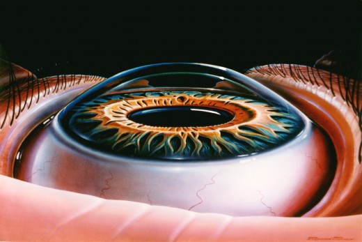
Many times, people go to the doctor without having a true understanding of how a part of their body works under healthy conditions. Thus, when a problem or ailment arises, or a doctor is too buy or in a hurry to see the next patient, a person may have a hard time understanding exactly what is wrong.
This article, using easy to understand terminology and illustrations, explains how the healthy eye works. For your next eye exam or doctor visit, you will have a perfect understanding of perhaps the most vital sense.
Other Works By The Author
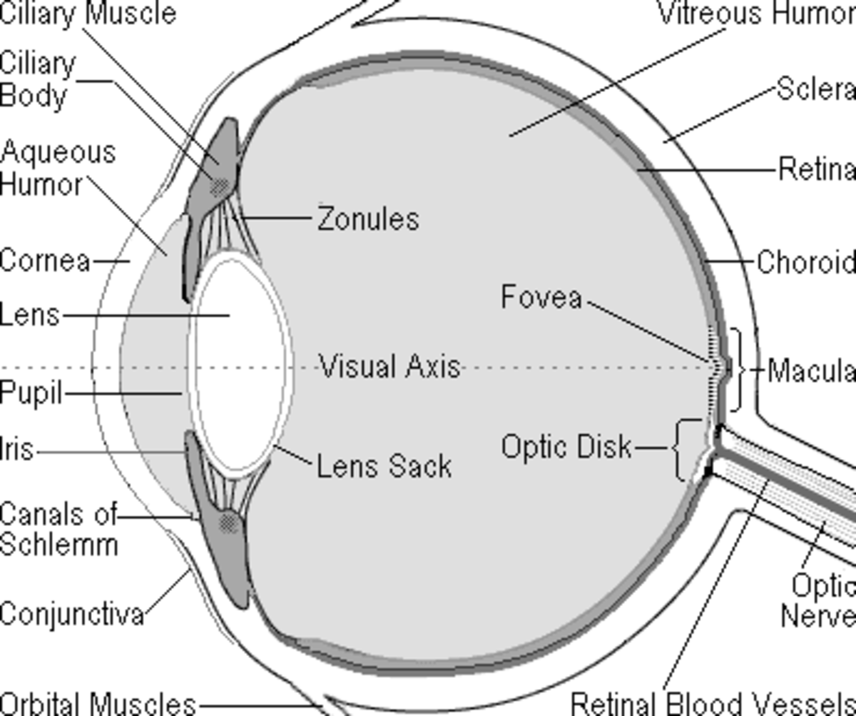
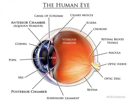
To truly understand how the function, it is perhaps easiest to think of the eye not dissimilar from a camera (not a digital camera, but those old camera from 1998 that actually used film!). Beginning with the world around you, images, in the form of light, enter the eye through the clear cornea, the outermost portion of the eye.
Most focusing occurs here, between the air and the curvature of this clear cornea. After passing through the cornea, light goes through the lens. Like a camera, light is further fine-focused through this lens.
Consisting of four layers, the lens is flexible and can change shape to better focus light entering the eye. The ciliary body, which encircles the lens, contain muscles which change the shape of the lens to focus light. From the lens, focused light then travels back to the retina.
On the back wall of the eye is the retina - a layer of cells which absorb light, much like the film near the back of a camera. The retina is an amazing collection of specialized living cells, which record moving images from the outside world - transforming light into nerve impulses.
The specialized cells contained in the retina are called rod and cone cells. These cells are nourished by a complex vascular structure beneath them, called the choroid (much like the root system of a tree).
Your clearest vision occurs at the center of the retina, in a very small area called the macula. The macula has a very high concentration of cone cells.
After being absorbed by rod and cone cells, images, in the form of nerve impulses, travel out of the eye through the optic nerve, where it enters the brain.
Another very important part of your eye is an area called the angle, located just inside the front of the eye near the outer edge of the colored iris. At the angle, there is a constant inflow and outflow of nourishing clear aqueous fluid, which travels from behind the iris, through the pupil, and out an area called the trabeculum.
As can be seen, the eye is a marvel of both biology and physics, composed of hundred of vital parts and tissue. The next time you go to a doctor, you can be rest assured, you will possess the knowledge to fully understand what is exactly going on inside your eyes.
Click here and here, or on my profile, for tips on maintaining your most precious gift - the gift of sight.
Matthew Gordon is the author of The Thin Blue Line: An In-Depth Look at the Policing Practices of the Los Angeles Police Department &
To Live, To Think, To Hope - Inspirational Quotes by Helen Keller.
© Matthew Gordon, 2011
*This article is a simple explanation article, and should not be taken as medical fact. A doctor should be consulted for all medical related actions.


