A Vet's Guide to Understanding Canine Arthritis and Degenerative Joint Disease
According to the Arthritis Foundation, in the U. S., pet owners can anticipate that some form of arthritis is responsible for their dog's chronic pain. Additionally, one in five dogs develops some type of arthritis.
Given those grim statistics, what symptoms might alert owners that their dogs are suffering from arthritis rather than some other ailment?
In today's interview with Dr. Cathy Alinovi of Hoofstock Veterinary Services, we'll learn what arthritis and degenerative joint diseases are, how to spot the early symptoms, and what methods veterinarians use to make a diagnosis.
Question 1: What is arthritis?
Dr. Cathy: Arthritis is the inflammation of one or more joints. Joint (arth) plus swelling (itis), and pain and stiffness are the most common symptoms, which tend to worsen with age. This condition is often called OA (osteoarthritis) (osteo-bone).
Q2: How many types of arthritis are there, and how do they differ?
Dr. Cathy: Arthritis is classified as either non-inflammatory or inflammatory based on what’s in the joint fluid (the soft swelling around the joint).
Non-inflammatory
Non- inflammatory joint disease (arthritis) manifests in degenerative changes in the joint but no fever or systemic signs. Inflammatory changes, as well as systemic (whole body) signs, characterize inflammatory arthritis/joint disease.
Inflammatory
Inflammatory arthritis is further broken down into infectious or immune-mediated. Infectious joint disease occurs when organisms (usually bacteria) invade a joint and cause inflammation. Immune-mediated joint disease occurs when the immune system is out of balance and it can cause severe joint damage, which is often seen on x-rays (radiographs).
Canine Arthritis
Classification of Arthritis
| Systemic Signs
| Cytology
| X-Ray
| Example
|
|---|---|---|---|---|
Non-inflammatory
| none
| -osteophytes, flattened or irregular femoral head, shallow acetabulum, sclerosis of subchondral bone
| Hip dysplasia , OA
| |
Inflammatory
| Yes- Examples: Fever, ↑ WBC
| Staph, Strep Coliforms, etc..
| ||
Infectious
| Yes- Examples: Fever, ↑ WBC
| >40,000 WBC/ mm^3
| +/_ erosion or osteomyelitis swelling/effusion
| |
Immune mediated
| Yes- Examples: Fever, ↑ WBC
| 5000-10,000 WBC/mm^3
| Changes
| Rheumatoid
|
Erosive
| Intermittent
| No changes
| ||
Non-erosive
|
Key Definitions
Cytology: study of the cells in the joint fluid in this case.
Osteophyte: bone spur in the joint, causes pain.
Femoral head: top of the leg bone.
Acetabulum: part of the pelvis where the femoral head sits; the socket to the ball and socket joint.
Sclerosis: hardening.
Subchondral: the layer right underneath the cartilage (chondro).
Peri: around.
Fibrosis: loss of stretch.
Q3: Which breeds are affected more by arthritis - small or large breeds - and why?
Dr. Cathy: On average, large dogs are affected more by arthritis due to their rapid growth phase as puppies, which results in an increased strain on the joints due to their size and weight. Rheumatoid arthritis, which is a form of immune-mediated arthritis, affects smaller breeds more often. While septic arthritis is more common in male sporting breeds, it can be seen in dogs of any age.
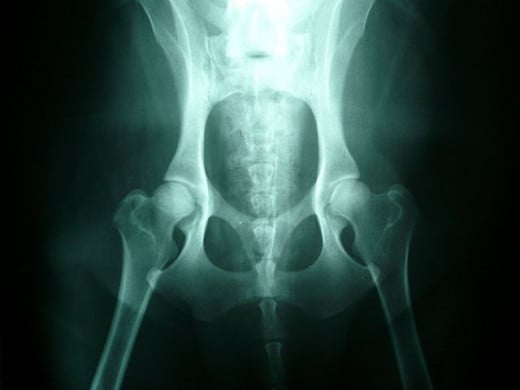
Normal Canine Hips
Q4: What forms of canine degenerative joint diseases are most common?
Dr. Cathy: DJD, which is degenerative joint disease or OA, also known as osteoarthritis and characterized by bone and joint inflammation, is a degenerative, progressive, and irreversible disease process affecting the synovial joints (like knees and elbows). Typically, progressive loss of articular cartilage, osteophyte formation and peri-articular fibrosis are seen with DJD/OA.
OA is classified as primary or secondary. Primary OA is an issue in cartilage that is associated with aging. Secondary OA is a consequence of an external event such as trauma, joint incongruity or misalignment, which negatively affects articular cartilage. Primary OA is most commonly seen in older dogs while secondary OA is the most common cause of clinical lameness in younger dogs.
DJD can present in many different ways. Here are some names for specific conditions, which ultimately still end up with DJD:
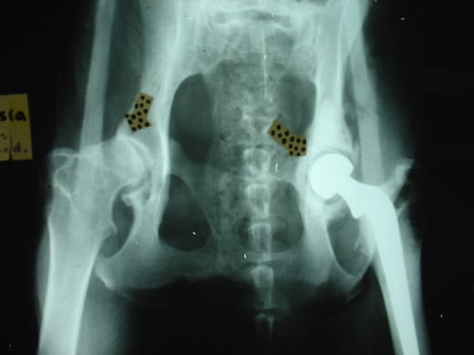
- Hip dysplasia: the leg bones do NOT form a nice, neat ball and socket joint with the pelvis. The result is looseness (laxity) in the joint. This can be quite painful.
- Joint trauma: includes articular fractures (tibial and femoral most typical,) which can be accompanied by ligament rupture and, subsequently, DJD.
- Patella luxation: when the patella (kneecap) does not stay in the groove (luxation). It is an inciting cause of stifle (knee) OA.
- Ruptured cruciate ligaments (torn ACL): most common cause of stifle OA, which is made worse if patella luxation is also present. These patients are usually non-weight bearing, but not always. Their condition worsens with exercise: they will be somewhat responsive to pain medications.
Anatomy - ACL
In the knee between the major leg bones (the femur and tibia), there are two ligaments that support the joint. These ligaments make an X with each other, and thus are referred to as cruciate, which is Latin for cross. In a torn ACL, the cruciate ligament that connects the back of the femur to the front of the tibia is torn. This is a very common injury for football players.
On examination, there is often joint swelling, pain on extension, crepitus (crunching), thickening of the joint on the inside of the knee (medial buttress), and the dog does not sit square. A veterinary exam will look for what is called a cranial drawer sign or cranial tibial thrust.
This test looks for movement of the lower part of the leg relative to the top, somewhat like a dresser drawer, in which the tibia slides forward under the femur at the knee (stifle) joint. In many big dogs, sedation is required to find the drawer sign as they have so much leg muscle they will not allow the painful movement when awake. The diagnosis is usually made based on x-rays. X-rays showing the presence of the “fat pad sign” are confirmatory. It looks like a dark line under the ligament.
Canine Hip Dysplasia
Q5: What signs would alert pet owners that their dogs might be suffering from arthritis?
Dr. Cathy: Any behavior change is reason to worry. These changes can include:
- Lameness
- Difficulty rising in the morning,
- Exercise intolerance/reluctance to move,
- Ataxia (wobbly walk),
- Pain/soreness
- Warmth or swelling over a joint
- Muscle atrophy (loss)
- Gait abnormality
Q6: What is the prognosis for dogs with arthritis?
Dr. Cathy: The prognosis depends on the type of arthritis and how early the disease is detected.
- Erosive: rarely resolves but remission is possible (semi-erosive polyarthritis of Greyhounds has a poor prognosis).
- Non-erosive: good to fair prognosis; may attempt to maintain in remission
- Septic: variable, depending on the extent of cartilage damage
- Torn ACL: OA progresses despite treatment. Dogs weighting less than 30 pounds are 80-90% subjectively normal within 3-6 weeks with diet changes, restricted activity (leash walks) and anti-inflammatory/pain medicine.
- Luxating patella: prognosis is good for milder cases, grades 2 & 3. The prognosis is guarded for more severe cases, grade 4. The older the condition is, the worse the prognosis.
- Neoplasia: depends on type of cancer, stage and treatment.
- Trauma: ligament damage will depend on the severity of the DJD. An articular fracture accompanied by DJD will typically result in the rupture of joint ligaments as well. These patients can have very painful outlooks.
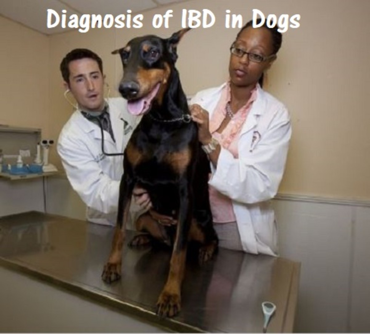
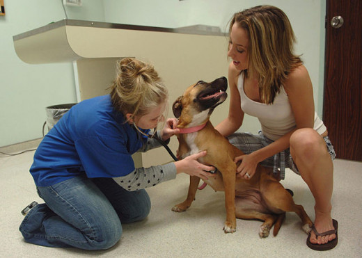
Q7: As a vet, what do you do during an arthritis examination?
I palpate joints feeling for crepitus (crunch) and watching for any painful response and/or joint swelling. I look for muscle imbalances and decreased range of motion in the joints through all angles of movement. I do a gait analysis where I watch a dog walk, trot and run. I evaluate the dog's neurologic state. Are the reflexes present? Are reflexes symmetrical? Are reflexes bright or dull? How is the dog's mental status?
Later, it may be necessary to run blood work, take radiographs (X-rays), or perhaps even perform a joint tap under sedation to collect fluid for analysis to look for infectious causes of the swollen joints. It is important to trim hair and nails to allow for proper motion and traction when moving during an exam.
All of this information plus information the pet parent can provide helps give us (the owner and myself) a full picture of the problem. With the problem well understood, we can look at how to treat the arthritic patient.
Chances are that if you suspect your dog is suffering from arthritis, he or she probably is. Getting an expert opinion of your pet's condition and starting any prescribed treatments as quickly as possible is the best way to make your canine friends more comfortable and help them to enjoy a better quality of life.
© 2013 Donna Cosmato

