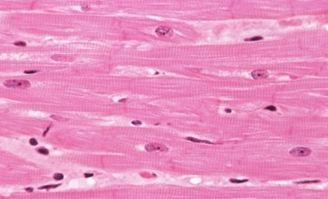
Anatomy of Smooth Muscles

Smooth muscle gets its name from the fact that it lacks the striations characteristics of cardiac and skeletal muscles and consists of small cells each with a single and central nucleus. Layers of smooth muscle cells line the walls of various organs and tubes in the body such as the
 muscle")
1. Blood vessels
2. Stomach
3. Intestines
4. Bladder
5. Airways
6. Uterus
7. Male and female reproductive tracts
AND the contractile function of smooth (visceral) muscle is not under voluntary control.
When made to contract the smooth muscle shortens allowing the contents within the organ to move along…such as in your digestive tract when you’ve just eaten something. Smooth muscle cells do lack the banding pattern found in cardiac and skeletal muscles and they receive neural innervation (supply of nervous energy or of nerve stimulation sent to a part) from the autonomic neural system. Contraction taking place in a smooth muscle is controlled by hormones and other chemical signals.
Smooth muscle is also divided into two sub-groups: the single unit and the multi unit smooth muscle.
Within a single unit smooth muscle tissue, the autonomic (involuntary) nervous system innervates a single cell within a sheet/bundle and the action potential is propagated by gap junctions (which allows for communication between cells) to neighbouring cells and causes the whole bundle/sheet to contract as a syncytium (a group of cells in which the cytoplasm of one cell is continuous with that of adjoining cells, resulting in a multinucleate unit).
Most of the smooth muscle found in the body is of the single unit variety however, multi unit smooth muscle tissue can be found in the trachea, iris of the eye and large elastic arteries. Multi unit smooth muscle tissue innervate individual cells therefore they allow for fine control and gradual responses.
A few pointers of smooth (visceral) muscle cells:
- each have one, single placed nuclei
- shorten on contraction
- are able to undergo cell division and regenerate after damage
- don't have sarcomeres nor do they have myofibrils
- have actin and myosin filaments that crisscross the cells and are inserted into the cell membrane at anchorage points
- are linked structurally and electrically into functional units
More articles on your muscular system
- Overview Of Muscle Types
Muscular system Indeed, rippling muscles of a weight lifter is often the first thing that may come to your mind when you think of the word muscle. The word muscle is actually derived of the Latin word... - Anatomy of Skeletal Muscles
Skeletal muscles are associated with movements of the body and these movements are the results of the unique characteristics of skeletal muscle cells. In the previous article we looked at the three types... - Anatomy of Cardiac Muscles
Cardiac muscles is a type of muscle found only in the walls of the heart and is responsible for pumping blood throughout the body - the function being transportation and the transportation vehicle being...


