Cells Of The Nervous System

Until later in the 1800s, the best microsope revealed very little about how the brain works. Only through special staining techniques could the tiny cells or neurons be seen. It was difficult to see each from the area that surrounded them.
Scientists noticed thin fibers between one neuron and another, however they could not distinguish where each fiber stopped or the next cell began or if all of them merged together.
In the later 1800s, Santiago Ramon y Cajal proved that a single neuron does not merge with other neurons or cells. A tiny gap separates the end of one neuron's fibers from the end of another neuron.
It's easy to understand why it was once believed that the cells of the brain worked as one big unit. We think of things working together much like a football team. It makes more sense that a neuron would be a part of a bigger working unit. We now know that this simply isn't the case.
The adult brain of a human contains an estimated 100 billion neurons. All of these neurons or cells are individuals that combine in effort to coordinate an organized behavior.
Neurons and Glia
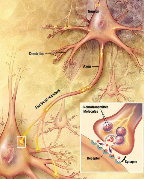
The nervous system consists of two types of cells, glia and neurons. Neurons are cells that receive information then pass it on to other cells. These are also what people call nerve cells. There are many types of glia and they serve many different functions. Glia cells cannot transmit information in long distances like neurons can, but they can exchange chemicals with neurons beside them.
Animal Cell Structures
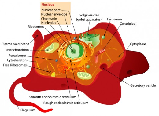
Every cell is surrounded by a membrane. A membrane is a structure that separates the inner and outer sides of the cell. It is made up of two laters of fat molecules that float freely around each other. Tiny uncharged chemicals, such as H2O or oxygen, move freely across the membrane. Some charged ions, such as potassium, choride, or sodium can cross through protein channels which are tiny openings. Most chemicals cannotcross through the membrane.
All animals cells have a nucleus except red blood cells. The nuceus is what contains the chromosomes. A mitochondrion is the structure where the cell does metabolic activities which gives the energy needed for the cell to do all its other activities. Mitochondria must have oxygen and fuel to function. Ribosomes are the areas which the cell synthesizes new protein molecules. Proteins provide what the cell needs to develop different chemical reactions. Some ribosomes freely float around in the cell while others are attached to the endoplasmic reticulum (a network of thin tubes that carry new synthesized proteins to other areas.
Neuron Structures
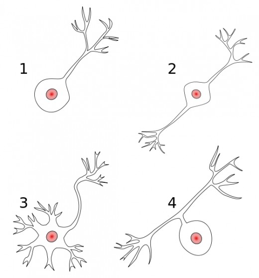
A neuron has a membrane, a nucleus, mitochondria, ribosomes and other structures found in animals cells. The shape of the neuron is distinctive.
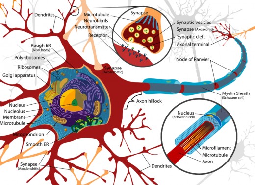
The majority of neurons have a cell body, an axon, dendrites, and presynaptic terminals. A motor neuron gets excitation from other neurons and produces impulses from its soma in the spinal cord to gland cells or muscles. Its dendrites go into the soma while the axon exits from it.
A sensory neuron has a specialized end and is very sensitive to a particular type of stimulation, such as information from skin cells. Different types of sensory neurons have different structures.
The dendrites are fibers that branch and get more narrow as they extend from the cell body toward periphery. The dendrites make up the information-receiving pole of the neuron. The dendrite's surface is laced with specialized synaptic receptors, where the dendrite receives information from other neurons. The bigger the surface of the neuron, the more information it can receive. A few dendrite branch out widely and have a much larger surface area. Some also have dendrite spines which are shorter outgrowths that make the surface area larger for synapses.
The cell body, or the soma, contains the the nucleus, mitochondria, ribosomes, and a few other structures found in cells. Most of the metabolic action takes place in soma. As with the dendrites, the soma is covered with synapses on its surface in many neurons.
The axon is a thin fiber of continuous diameter. The axon sends the information of the neurons. It conveys an impulse toward other neurons or a muscle or gland. Many verebrate axons have an insulating substance called a myelin sheath that covers it. The axons of invertebrates do not have this.
An axon contains many branches, each swells up at it's end, making a presynaptic terminal, also known as a bouton or end bulb. This is the tip where the axon releases chemicals that go through the junction between neurons (from one to another). Since synthesizing these chemicals extract a lot of energy, the prosynaptic terminals have quite a few mitochondria.
A neuron can have different numbers of dendrites, but can only have one axon, which may have many branches. Some axons are very long. For instance, specific axons from a human's spinal cord runs all the way down to the feet.
A local neuron is very small with no axon, in some cases it may contain a very small axon. It can carry information only to other neurons adjacent to it.
An afferent axon carries information into a structure. An efferent axon takes information away from the structure. All sensory neurons are afferent to the rest of the nervous system. All motor neurons are efferent from the nervous system. One way to remember the difference of these two terms (afferent and efferent) is their beginning letters "a" and "e"- efferent starts with e, remember exit, and affarent starts with a, remember add.
If a cell's axons and dendrites are completely inside a single structure it is an interneuron or intrinsic neuron of that structure.
Neurons come in all shapes and sizes and vary in fuction. The shape of a neuron determines it's correlation with other neurons. Therefore, it determines its functions of the nervous sytem. The wider the branches, the more connections with other neurons.
Glia
Glia's functions are not understood completely yet. One special type of glia, the astrocyte, is star-shaped and wraps around the presynaptic terminals of several axons, possibly a work in unison. An astrocyte helps synchronize the workings of axons by taking up chemicals released by those axons then later releasing those same chemicals back to the axons. This enables them to send information in waves. Astrocytes aslo function by removing waste material, such as dead neurons.
Other specialized types of glia are Oligodendrocytes (in the brain and spinal cord), and Schwann cells (in the periphery of the body). Oligondendrocytes and Schwann cells build the myelin sheaths that surround and insulate specific vertebrate axons.
Radial Glia, an astrocyte, guide the movement of neurons and the growth of their axons and dendrites at the stage of embryonic development. Schwann cells guide regenerated axons to the targeted location after damage.


