Development Of The Vertebrate Brain


At two weeks old the human embryo begins to form its central nervous system.
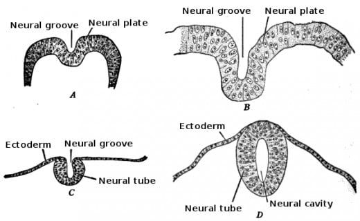
The dorsal surface thickens first, then thin, long lips raise up, curl, and form a neural tube surrounding a fluid-filled cavity. The tube then sinks beneath the surface of the skin, as the front end enlarges and separates into the midbrain, hindbrain, and the forebrain. What is left over afterwards becomes the spinal cord. The fluid-filled cavity inside the neural tube turns into the central canal of the spinal cord and the 4 ventricles of the human brain.
The process is similar in about all vertebrates. The only real difference is the amount of time to develop, and the size.
The human brain weights about 350 grams at birth. Specific areas of the forebrain are under-developed for the first few weeks, which is indicated by their low levels of glucose use. The developmental stage is very rapid. Certain areas of the brain that are almost dormant at birth show adult patterns of arousal within 8 months. At the first year milestone, the human brain weighs 1000 grams, which is not far from the adult brain weight of 1200 to 1300 grams.

Development of Neurons
With the development of the nervous system comes the production of neurons. Neuroscientists differentiate four distinct and major stages in the development of neurons:
- Proliferation is the production of new cells. Very early in the developmental stage, cells lining the ventricles of the brain start dividing. A few of the new cells stay where they are formed, and continue to divide and redivide.
- Migration is the moving of cells toward their eventual destination in the brain. This process requires a perfect chemical environment. Any interference with migration or proliferation can result in cognitive deficits to mental retardation.
- Differentiates is the forming of axon and dendrites that provide its unusual or distinctive shape. The axon grows prior to the dendrites. It actually grows during the migration of the neuron. A few neurons have an axon growing anterior to them like a tail. When the neuron gets to its final destination, dendrites will begin to form, extremely slow at first. Most of the dendrite growth happens much later in development, when incoming axons are about to arrive.
- Myelinate is when glia cells produce the insulating sheaths. In humans myelin forms first in the spinal cord, then in the hind brain, mid brain and then the fore brain. Very different from migration and proliferation, myelination continues for years and years. Quite a few number of myelin is still forming at the age of 20, and in a few brain areas it continues for decades.
Development of The Neural Tube
Maturing of the brain doesn't only need development of neurons but also the organization of brain areas. Scientist discovered what happens as the brain grows, at the microscopic level, by staining and photographing the living brain of an infant mouse and then photographing that same area later in the developmental stage. This experiment proved that the brain adds subdivisions during at least the first 3 weeks of life.
The brain develops by both expanding old areas and by adding new ones. This is true in all species of animals with large brains.

Determinants of Neuron Survival
The right number of neurons for each specific area of the central nervous system isn't easy, it's complicated. To be useful, each neuron has to receive axons from the correct source and send its own axons to a specific cell in the right area. The vast number of areas do not develop at the same time or rate, so at times the neurons in one specific area may develop prior to any incoming axons have arrived or before any receptive site becomes available for its own axons. Ironically, we find no left-over neurons which failed to make its connection.
When a neuron of the synthetic nervous system develops a synapse on an organ muscle, the muscle sends a protein called nerve growth factor or NGF. This protein promotes the growth and survival of the axon. An axon that does not receive the right amount of NGF cannot function properly and its cell body dies. Each neuron begins with an innate program of a suicide mission. If its axon does not make the connection with an appropriate post synaptic cell by a specific age, the neuron will kill itself. The process through which this neuron kills itself is called apoptosis (an innate programmed mechanism of the death of a cell).
Apoptosis differs from necrosis. Necrosis is when the death is caused by injury or a toxic substance. NGF cancels the program for apoptosis. This protein NGF simply gives a much needed protein that tells the postsynaptic cell not to do the suicide mission.
Nerve growth factor or NGF is a neurotrophin. It is a chemical that endorses the survival and promotes the activity of neurons. These neurotrophins are useful in many ways.
Early in development, they cause specific axons to survive and thrive instead of submitting to apoptosis. Different neurotrophins are available and active for different types of axons.
Much later, neurotrophins are secreted by neurons because of new experiences. This increases the branching of incoming axons and facilitates the system and mechanisms that store memories.
Neurotrophins decrease pain and increase regrowth of damaged axons in response to a serous system injury.
Scientists are hopeful that someday they will be able to utilize the power of neurotrophins and use it to relieve certain diseases that attack the nervous system.
All areas of the developing nervous system, in the beginning of development, overproduce neurons, sometimes three times what is needed to survive into adulthood. Each area of the brain has a time of massive cell death. This loss of cell death doesn't mean there's something horribly wrong, it is actually a normal part of the developmental stage.
One possible explanation for producing way more neurons that needed is that the excess of neurons serves to help in error correcting. If some axons don't reach specific targes, others will. Another reason for over-producing neurons is simple; there is no possible way to determine how many fibers a leg muscle or arm muscle will have, therefore the spinal cord will produce an ample supply of neurons and later get rid of the excess.

Credits:
- Levi-Montal-cini, 1987
- Gotz et al., 1993; Mendell, 1995
- Kesslak, So, Choi, Cotman, & Gomez-Pinilla, 1998
- Beck et al., 1995
- Finlay & Pallas, 1989


