Human Reproductive System

Male Reproductive System
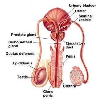

Male Reproductive System
It consists of a pair of testes, accessory glands and a system of ducts. Testes secrete the male sex hormone called 'Testosterone' and produces spermatozoa or sperms (haploid).
Inside each testis, several lobules are present. Each lobule has several tubules called 'Seminiferous Tubules'. The epithelial cells lining these tubules are called Germinal epithelium which undergoes several mitotic divisions and one meiotic division to produce spermatozoa and are released into the lumen of the tubule. After their release, spermatozoa enter a duct. This is called Vas efferentia and then into epididymis in which sperms are stored temporarily. From the epididymis, sperms move through Vas deferens. Later they enter into the urethra and expelled out of the body.
The accessory glands include one prostate gland, two seminal vesicles and two couper's glands. Secretions of these glands are called semen. These secretion provide nutrients for the sperms and also provide the fluid medium for the movement of sperms.
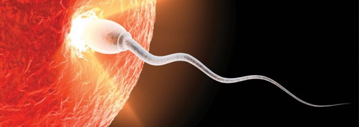
Structure of human sperm
Human sperms are minute, microscopic and motile and consists of an oval head piece, a neck, a middle piece and a long end piece or tail. Head piece consists of a large haploid nucleus and an Acrosome, a sac like structure, which helps in fertilization. The neck is short.
Middle piece has several mitochondria, which produce energy required for the movements of sperms. Tail piece helps in the swimming of sperms to reach the ovum during fertilization.
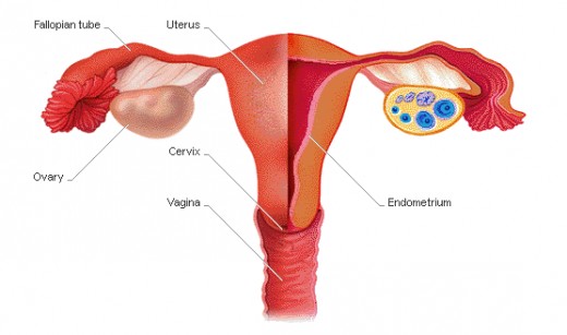
Female reproductive system
It consists of a pair of ovaries, fallopian tubes, uterus and vagina. Ovaries are the lobular structures present in the abdominal cavity. They are held in position by ligaments. Each ovary has several lobes called Graffian follicles.
From each follicle develops one ovum by meiotic divisions. Every time, only one follicle matures and releases one ovum into body cavity. The ruptured follicle is called Corpus Luteum and it secretes female sex hormones - oestrogen and progesterone.
A pair of fallopian tubes are present on either side of the uterus. They extend from the ovary and uterus. The anterior end of the tube is funnel like and ciliated. It helps in collecting the ova released into the body cavity and transport them into the uterus. Uterus is a single glandular sac with thick walls.
It facilitates the development of fertilized egg into foetus Vagina is a muscular tube into which uterus opens. Vagina opens out through female genital opening.
Ovulation and Menstrual Cycle
In females, formation of ova from the primary oocyte begins on the onset of puberty. The discharge of ovum from the ovarian follicle is called ovulation.
In males, the production of sperms occurs continuously throughout the life. But in females, the production of ova is not continuous and occurs once in 28 to 30 days. During this period, the female reproductive system undergoes a lot of structural changes in anticipation of fertilization and pregnancy. If fertilization does not take place, the released ovum degenerates in 24 hours. Once again, changes are repeated to produce another ovum. This cycle of changes in the female reproductive system called menstrual cycle.
Menstrual cycle has two phases - Proliferative and Secretory phases.
Proliferative phase
This phase occurs during the first 12 to 14 days of the cycle. During this period, the cells of the follicle, fallopian tube, uterus etc. increase in size. It is during this phase the follicle matures and releases the ovum. Growth of the tissues occurs in anticipation of fertilization and pregnancy.
Secretory Phase
This is the second half of the menstrual cycle. During this phase, the ruptured follicle is converted into corpus Luteum. The uterine walls are thickened so as to receive the zygote. When fertilization does not occur, the cells of Corpus Luteum and the uterine walls are expelled out along with some amount of blood.
The menstrual cycle is under the control of hormones secreted by the pituitary (follicle stimulating hormone, Lutenizing hormone and Prolactin) and those secreted by the ovaries (Oestrogen and Progesteron).
Fertilization and implantation
Fertilization is internal. Sperm reaches the ovum in the fallopian tube. During fertilization, chromosomes of the ovum and sperm make up into pairs and the resulting cell is called zygote. The diploid zygote undergoes several mitotic divisions and develops into embryo. The embryo is called as Blastocyst. This encloses blastocoel. The blastocyst slowly moves through the tube and enters the uterus. By this time, the uterine wall becomes thick, glandular and sticky. Blastocyst attaches with the wall and is slowly embedded in it. This is called implantation. Further development of the blastocyst occurs in the uterus only
Embryonic development
The growing embryo forms two membraneschorion and Amnion.
Chorion
It establishes connection with the walls of the uterus and aids in the proper supply of nutrients to the growing embryo. It also removes the wastes from the embryo.
Amnion
Amnion forms a sac like structure around the embryo and the space is filled with amniotic fluid. This fluid protects the embryo against mechanical shocks. As the embryo grows, the amnion also grows in size. It retains till the child is born.
Cells of the embryo differentiate into 3 layers Ectoderm, Endoderm and mesoderm. Cells in these layers proliferate and differentiate into various organs of the body.
Placenta
It is formed by the cells of the embryo and the mother. It is formed around 10-12 weeks of pregnancy. Placenta is an important structure for the nutrition of the embryo. Blood vessels of both mother and embryo are present in the placenta. The embryo receives the required nutrients, oxygen etc. from the mother through the blood vessels present in the placenta.
During this period, the tough cord called 'Umblical Cord' is also formed by the embryo, which is connected to the walls of the uterus through the placenta.
From the 3rd month of pregnancy embryo is called Foetus. The total time required for the embryonic and foetal development is called gestation period which is usually about 40 weeks. After this, the foetus is expelled from the uterus. This is very complicated process. The foetal hormones are produced and stimulate the contractions of the walls of uterus. The contractions are called "labour pains" and help in the expulsion of the foetus. The amnion ruptures first releasing the amniotic fluid out. Then the foetus is expelled out. Later the placenta is separated from the uterine walls and expelled out. But the foetus is still attached to the uterus of the mother by the umblical cord, which is separated by the doctor.