Introduction to Microtubules - Another way to try and stop cancers
Introduction to Microtubules
Hello and welcome to my article on cytoskeletal structures in eukaryotic cells called microtubules.
This article will consist of the properties and functions of microtubules, the role of tubulins as assembly blocks of microtubules, microtubule organising centre (MTOC), microtubules associated proteins (Maps), dynamic instability of microtubules and microtubule drugs. At the bottom of this article you will find the role of microtubules in removal of cancerous cells and microtubule drugs.
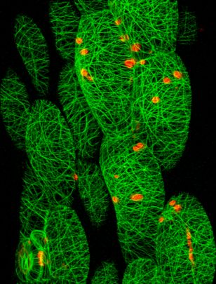
Microtubules (MTs) are one of three main cytoskeletal structures in eukaryotic cells. MTs act as beams that provide mechanical support for the shape of cells. MTs act as tracks long which molecular motors move organelles from one part of the cells to another and separate chromosomes during mitosis. The three main cytoskeletal structures in eukaryotic cells include Actin filaments (5-9nm diameter), intermediate filaments (10nm diameter) and microtubules (24nm diameter).
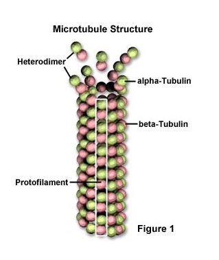
A microtubule has two different types of subunits known as alpha tubulins and beta tubulins which contain and plus end and a minus end. The structure of a microtubule and its subunits is hollow, cylindrical, polar and is made up of tubulins. MTs are polarised polymers of tubulins meaning that there is a plus end and a minus end to the molecule. Alpha and beta heterodimers bind head-to-tail along protofilaments. Alpha and beta subunits unite to form a heterodimer (8nm in length) and rows of heterodimers form protofilaments. Protofilaments are slightly out of step forming a helical arrangement. Usually a microtubule is made up of 13 linear rows of protein molecules. Each tubulin binds to GTP which will be explained further along this article.
MTs are polar filaments because the two ends have different structures and different properties, a minus end and a plus end. Microtubules are typically made up of 13 linear tows of protofilaments although MTs can also be 10-16 protofilament rows.
Microtubule functions include providing structure acting as an internal skeleton, as a polarised track for microtubules motors including kinesins and dyneins that drive movements within cells. Microtubules motors segregate chromosomes in mitosis, give the cell polarity and produce polarised organisation of organelles, regulate cell motility and cell division.
Microtubules are crucial in the organisation of chromosomes and cell organelles in interphase and mitosis.
Microtubules are abundant in neurons and the brain is the best source of tubulin.
MTs are organised in a cell in a specific manner to function properly. The microtubules organizing centre (MTOC) is located in the centre of the cell. Centrosome and centrioles are specific to MTs and are crucial to mitosis.
A centrosome is a microtubules organizing centre (MTOC) MTOC’s control where microtubules are formed. Centrioles within centrosome become basal bodies which are nucleation centres for cilia and flagella. Basal Bodes are Nucleation centres for cilia and flagella.
Microtubule associated proteins (Maps) control microtubules assembly and many stabilizing Maps bind to outer surface of protofilaments.
Microtubule ends exhibit dynamic instability. Dynamic instability in microtubule allows the quicker growth on the plus end (terminated by beta-subunit) where as a microtubules will grow very slowly on the minus end (terminated by the alpha subunit). Plus ends will sometimes switch depending on which phase of growth the cell is currently in i.e. slow growth or rapid shrinkage.
Dynamic instability is important because it explain how microtubules could reassemble into different structures during the cell cycle or during development. Dynamic instability normally only involves the plus ends as the minus ends are bound to MTOC and thus stabilised. Plus ends will also grow faster and are more dynamic than minus ends.
Research into microtubules dynamic in living cells is done through microinjection of labelled tubulins by staining with vital dyes then using digital-imaging fluorescence microscopy.
Microtubule growth occurs through GTP – stabilised dimers that add to the growing plus ends. Later these GTP dimmers can be dephosphorylated to GDP, thus microtubules are polarised. Dynamic properties include temperature decrease or pressure increase changes can depolarised the dimers and cause the breakdown of MTs. Plus ends of the microtubules are rapidly growing and collapsing and the minus ends are more stable. Microtubule ends change conformation between growth and shortening phases. GTP hydrolysis cycle is coupled to structural changes in the microtubule.
Microtubule Drugs:
Bind tubulin dimer and blocks assembly, colchicines, nocodazole, vinblastine, podophilotoxin, vineristine.
Binds dimer in microtubule lattice and stabilises microtubules.
Certain chemicals can influence MT stability. Colchcine bind to a dimer and prevents further addition, this property halts cell division at prophase and is known as an anti-cancer agent. Taxol from the yew tree is used in cancer treatment as it stops MT from breaking down and also prevents cell division. Other drugs such as vinblastine Vincristine and griseofluvin exhibit similar properties.
Research into microtubules can be used to treat cancers because MTs are crucial to mitosis (cell division). Some cancerous cells continue to divide constantly as the gene for cell division is being over expressed and not regulated. A drug that prevents microtubules from forming or does not allow microtubules to function would stop cell division and would thus prevent the cancer from growing (and may eventually die due to no less structural support) and easier to manage.
Share or vote this article if you find it interesting and post anything else you would like to share.
If you find my Hub interesting don't hesitate in leaving a comment, I would really appreciate it.
Thanks
Mike
