Modern Microscopes


History of the Microscope
Galileo Galilei placed two glass lenses inside a cylinder and viewed the patterns of an insect's eyes in the early seventeenth century. He was one of the first to view and record an observation with a microscope.
During mid-century, Robert Hooke viewed a thinly sliced cork from a tree and saw tiny compartments in which he named cellulae. This became the origin of the term "cell". What Hooke actually saw was dead plant cell walls. Hooke or nobody else at that time didn't think of cells as dead. In fact, they didn't realize cells could be living.
In the 1600s a shop owner named Antony Van Leeuwenhoek made lenses that allowed him to view sperm, bacterium, and protists.
From the 1820s through 1840 better lenses allowed better understanding of how cells work and divide.

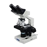
Compound Light Microscope
As previously used, most microscopes still use light rays to produce images. Each wavelength equals the distance from one wave's peak to the peak of the wave behind it. Light travels in waves as it moves from one place to another.
With a compound light microscope, two or more sets of lenses made of glass bend light waves that passes through a specimen, usually a cell, in a way that makes the view much larger.
Cells can be viewed under a microscope when they are thin enough for light to pass through them, however, most cells are without color and look dense. In these cases colored dye is used to stain cells so they can be visible.The most effective microscopes enlarge cells up to 2,000 times. More than that the cells visibility becomes blurry. This is caused by the parts being so small they are less than 1/2 of a wavelength of light, making them too small to make light bend.
Electron Microscope
Electron microscopes work by magnetic lenses which bend and focus beams of electrons. Glass lenses will not bend or focus electrons. Electrons travel in wavelengths that are about 100,000 times shorter than visible light wavelengths. Therefore, electron microscopes can bring objects into focus 100,000 times smaller than a compound light microscope.
Transmission Electron MIcroscope
Electrons pass through a specimen and are targeted into an image of the sample's internal parts in a Transmission electron microscope. In scanning electron microscopes, a beam of electrons travel back and forth over a specimen than has a thin layer of metal. The metal produces its own electrons and x-rays, which converts into a view or image of the surface. These images have amazing details.
- 6 Lifeforms Under A Microscope
A sort of micro-organism guide for dummies written in layman terms. Basically what I observed and learned using my microscope. Showcases 6 different lifeforms with video that I identified. - Parts and Functions of a Light Microscope (Part II)
A primitive microscope was invented in 1590 in Middelburg, Netherlands, by the eyeglass makers Hans Lippershey, Zacharias Jansen and his father Hans Jansen. Further, Galileo Galilei improved the instrument by using a set of aligned lenses and...


