Nervous System Review

Organization of the Nervous System
A Little Review:
The vertebrate nervous system consists of:
- A brain, a spinal cord, and a set of peripheral nerves.
The central nervous system or CNS
- Contains the brain and spinal cord.
The peripheral nervous system or PNS
- Consists of the cranial and spinal nerves that connect the CNS to all tissues.
A neuron
- Is an excitable cell that communicates via an axon.
A nerve
- Is a bundle of axons that carries information.
The afferent part of the PNS
- Carries sensory information to the CNS.
The efferent part of the PNS
- Carries information from the CNS to muscles and glands
The Afferent Pathways
Carries information to the CNS
- Stimuli from the sensory systems, pain and limb position move through afferent pathways.We have awareness of these stimuli. (Conscious)
- Information such as blood pressure, deep body temperature, and blood oxygen supply are also transmitted though afferent pathways.We are not aware of this information.(Unconscious)
The Efferent Pathway
Carries information from the CNS to the muscles and glands of the body.
Efferent pathways can be divided into two divisions:
- The voluntary division, the somatic division. Which executes conscious movements
- The involuntary, or autonomic division. Which controls physiological functions
Additional Info:
The CNS also receives chemical information from hormones circulating in the blood, or from neurohormones released by neurons into the extracellular fluids of the brain.
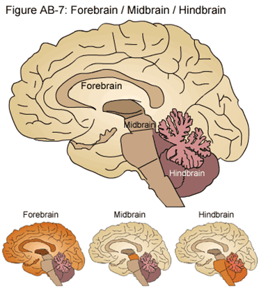
The Brain: Basic Structure
The Hindbrain
Consists of the Medulla, the Pons….
- Control physiological functions, such as breathing and swallowing.
- All information traveling from the spinal cord must pass through the pons and medulla.
And the Cerebellum
- Were muscle control is coordinated.
The Midbrain
Houses structures that process visual and auditory information.
Together the hindbrain and midbrain are known as the brain stem.
Note: Most sources do not include the cerebellum as part of the brain stem, but some do.
The Forebrain
Consists of the Diencephalon and the Telencephalon
Diencephalon
Consists of the Thalamus and…
- Final relay station for sensory information
Hypothalamus
- Regulates physiological functions such as hunger and thirst
Telencephalon
Cerebrum which consists of two cerebral hemispheres.
- In humans, this is the largest brain region. It is involved in sensory perception, learning, memory, and behavior.
Limbic system
- Primitive region involved in emotions and memory
- Hippocampus
- Amygdala

The Limbic System
Structures in primitive regions of the telencephalon form the limbic system.
- This is responsible for basic physiological drives, instincts, and emotions.
Consists of two parts:
Amygdala
- Involved in fear and fear memory
Hippocampus
- Transfers short-term memory to long-term memory
The Cerebrum
Is the largest part of the brain in mammals.
It is separated into two hemispheres connected by the corpus callosum:
- Cerebral hemispheres
The hemispheres are covered by a ‘sheet’ of gray matter that is convoluted to fit into the skull
-Cerebral cortex
- It consists of Gyri: (sing. gyrus): Ridges
- And Sulci: (sing. sulcus): Valleys
The Cerebrum Is divided into four main lobes.
- Temporal
- Frontal
- Parietal
- Occipital
Each lobe has a specific function

The Temporal Lobe
Receives and processes auditory information.
Areas of the temporal lobe also involve:
- Identification of objects
- Object naming
- Recognition of objects and faces.
Agnosia
- Disorder of the temporal lobe that causes someone to be aware of an object or stimulus, but can not identify or name it.
The Frontal Lobe
Located in front of the central sulcus
- Which separates the frontal and parietal lobes.
Contains the primary motor cortex.
- The neurons in this region control muscles in specific parts of the body.
- Areas with fine motor control have disproportionate representation. (Such as the hands and the face.)
Additional areas involve
Planning
Personality
- Phineas Gage, a nineteenth-century railroad worker whose ‘nice guy’ personality was transformed by an explosion that blew a tamping iron through his brain
The Parietal Lobe
Located behind the central sulcus.
- Remember this separates the frontal and parietal lobes.
Contains the primary somatosensory cortex.
- This area receives touch and pressure information.
- Just like the motor cortex, the whole body surface can be mapped onto the primary somatosensory cortex.
- Areas of the body that have a high density of mechanoreceptors and are capable of making fine discrimination in touch have a large representation. (Fingers and face.)
The Occipital Lobe
Receives and processes visual information
Additional areas involve:
- Making sense of the visual world
- Translating visual experience into language
Cerebellum
The cerebellum is important for coordination and error checking during motor, perceptual, and cognitive functions
- It is also involved in learning and remembering motor skills
The Brain
The Spinal Cord
Its Job:
Conducts information between brain and organs.
- Integrates information coming from PNS.
- Responds by issuing motor commands.
The Anatomy
Gray matter
- Is located in the center
- Contains cell bodies of spinal neurons, dendrites and unmyelinated axons.
White matter
- Surrounds gray matter
- Contains myelinated axons that conduct information up and down the spinal cord.
Spinal nerves
Extend from the spinal cord at intervals.
Each one has two roots
- Dorsal Root
- Connecting to the dorsal horn of the gray matter
- This is were afferent (sensory) axons enter the spinal cord.
- Ventral Root
- Connecting to the ventral horn.
- This is were efferent (motor) axons leave the spinal cord
The Spinal Reflex Arc
Sometimes afferent information can convert to efferent activity without the brain.
- This is called a spinal reflex.
A monosynaptic spinal reflex. (The simplest kind)
Involves only two neurons and one synapse
- The knee-jerk reflex is an example
Stretch receptors send action potentials through the dorsal horn to the ventral horn
- via sensory neuron axons.
In the grey matter the afferent (sensory) neurons synapse with efferent (motor) neurons in the ventral horn.
- Action potentials are sent along the efferent axons to leg muscles, causing contraction.
The Spinal Cord
Information Processing: The Autonomic Nervous System (ANS)
It Controls involuntary functions
Comprised of two divisions:
Sympathetic and Parasympathetic.
- They work in opposition to each other, one causes an increase in activity, the other a decrease.
- They each have different neurotransmitters and anatomy
Sympathetic division is responsible for:
- Increasing heart rate, blood pressure, and cardiac output.
- Decreasing digestion.
- Remember the “flight-or-fight” response?
Parasympathetic division is responsible for:
- Decreasing the heart and blood pressure.
- Increasing digestion
ANS
Both divisions of the autonomic nervous system are efferent pathways.
They begins with a neuron whose cell body is in the brain stem or spinal cord
- They are called cholinergic neuron because they use acetylcholine as a neurotransmitter.
- These cells are also called preganglionic neurons because the second neuron in the pathway with which they synapse resides in a ganglion
- Which is a collection of neuronal cell bodies that is outside the CNS.
The second neuron is called a postganglionic neuron because its axon extends out from the ganglion.
- The axons of the postganglionic neurons end on the cells of the target organs.
Neurotransmitter Differences
Sympathetic postganglionic neurons are noradrenergic.
- They use norepinephrine as their neurotransmitter.
Parasympathetic postganglionic neurons are mostly cholinergic.
- They use acetylcholine as their neurotransmitter.
Anatomical Differences
Sympathetic division:
- Preganglionic neurons originate from the thoracic and lumbar regions of the spinal cord.
- Most of the sympathetic ganglia are lined up on either side of the spinal cord.
Parasympathetic division:
- Preganglionic neurons originate from the brain stem and the sacral region of the spinal cord.
- The parasympathetic ganglia are close to the target organs


 - More Effectively than Conventional Methods")
