The Human Circulatory System

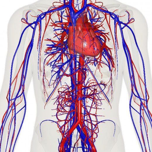
The circulatory system also known as the cardiovascular system consist of the heart, which pumps blood, and the blood vessels through which blood flows to the body.
THE HEART
The heart is a hollow, muscular organ situated between the two lungs in the mediastinum and rests on the diaphragm. The heart is about 14 centimeters long and 9 centimeter wide in an average adult. The heart is enclosed in a serous membrane called the pericardium. The pericardium consist of two portions, the fibrous pericardium, which is a dense connective tissue, surrounds a more delicate double layered sack, and the visceral pericardium which covers the heart. At the base of the heart, the visceral pericardium turns back on itself to become the parietal pericardium, which forms the inner lining of the fibrous pericardium. Parietal pericardium also lines the wall of the heart. the parietal and visceral layers of the pericardium is a space called the pericardial cavity, it contains a small volume of serous fluid which reduces friction between the pericardial membranes as the heart moves within them. Blood is supplied to the heart through the two branches of aorta known as the right and left coronary arteries. The heart must beat continually to supply blood to body tissues. For this to happen, myocardial cells require a constant supply of freshly oxygenated blood.
THE LAYERS OF THE HEART WALL
The wall of the heart is composed of three distinct layers.
(1) Pericardium: this is the outer layer of the heart wall, it consist of visceral layer of serous and fibro elastic and adipose tissue. It protects the heart by reducing friction.
(2) Myocardium: this is thick middle layer of the wall of the heart, it consist of cardiac muscle tissue that pumps blood out of the heart chambers.
(3) Endocardium: this is the inner layer of the heart wall, it consist of thin layer of endothelium and connective tissue, which contains elastic and collagenous fibers. It also contains some specialized cardiac muscle fibers called purkinje fibers.
CHAMBERS OF THE HEART
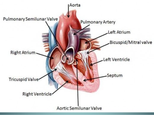
The heart is made up of four chambers. Two atria (left and right atrium) and two ventricles (right and left ventricles). The upper chamber atria has a thin wall and receives blood returning to the heart. An auricle are small earlike projections, it increases the capacity of the atrium slightly. The lower chamber, the ventricles receives blood from the atria and contract to force the blood out of the heart into the arteries. The atrium and ventricle are separated on the right side from their counterpart on the left by a septum. This separation allows blood from one side of the heart not to mix with blood on the other side of the heart. the tricuspid valve on the right and mitral valve on the right known as Atrioventricular valve makes sure that the blood flow one way between the atria and the ventricles.
THE RIGHT SIDE OF THE HEART
The right atrium receives blood from two large veins, the superior vena cava, and the inferior vena cave. In addition, a small, the coronary sinus drains blood into the right atrium from the myocardium of the heart itself. The tricuspid valve with three cusps lies between the right atrium and the right ventricle. This valve allows blood to move from the right atrium into the right ventricle and prevent back flow. A strong fibrous string, the chordae tendineae attaches to the cusps of the tricuspid valve on the ventricular side. It originate from small mounds of cardiac muscle tissue, the papillary muscle. The papillary muscle contracts as the ventricle contracts, and as the tricuspid valve closes this muscle pull on the chordae tendineae and prevents the cusps from swinging back into the atrium.
The right ventricle has a thinner muscular wall than the left ventricle. The right ventricle pumps blood a short distance to the lungs against a relatively low resistance to blood flow, while the left ventricle forces blood to all other part of the body against a much greater resistance to flow. When the muscular wall of the right ventricle contract, the blood inside its chamber is put under increasing pressure and the tricuspid valve closes passively. As this happens, the only exit for the blood is through the pulmonary trunk, which divides to form the right and left pulmonary arteries that leads to the lungs. At the base of the pulmonary trunk is a pulmonary valve with three cusps. The pulmonary valve allows blood to leave the right ventricle and prevent back flow into the ventricular chamber. The pulmonary and aortic valve are called semilunar because of the half-moon shape of the cusps.
LEFT SIDE OF THE HEART
The left atrium receives blood from the lungs through four pulmonary veins. Two from the right pulmonary veins and two from the left pulmonary veins. The interatrial septum separates the left atrium from the right atrium. Blood passes from the left atrium into the left ventricle through the mitral valve or bicuspid valve, which prevents blood from flowing back into the left atrium from the ventricle. The papillary muscles and chordae tendineae prevents the cusps of the mitral valve from swinging back into the left atrium during ventricular contraction. When the left ventricle contracts, the mitral valve closes and the only exit is through a large artery called the aorta. At its base is the aortic valve with three cusps. The aortic valve opens and allows blood to leave the left ventricle as it contracts. When the ventricular muscles relax, this valve closes and prevents blood from backing up into the ventricle.
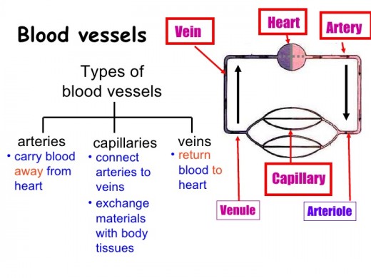
BLOOD VESSELS
The vessels of the circulatory system are aorta, artery, arterioles, capillaries, venules, veins, and vena cava.
ARTERIAL SYSTEM
The arterial system is made up of the aorta, arteries, and the arterioles. Three layers form aorta and arteries wall.
(1) Outer tunica adventitial: it is made up of connective tissue.
(2) Middle tunica midia: this is made up of smooth muscles.
(3) Inner tunica intima: it is made up of endothelia cells.
The aorta has a diameter of about 25mm, the arteries has a diameter of about 4mm while the arterioles has the diameter of about 30Nm(nanometer). The arterioles are continued as the capillaries, which are small, thin wall vessels with diameter of about 8Nm. the capillaries is very important because the exchange of material between the blood and tissues occurs at the capillary level.
VENOUS SYSTEM
From the capillaries, the venous system starts and it includes the venules, veins and the vena cava. Capillaries ends in the venules and these venules are small vessels with thin muscular wall than the arterioles. The diameter of the venules is about 20Nm (nanometer). At a given time, large quantity of blood is held in the venule, and therefore the venules are called capacitance vessels. the venules are continued as veins which have a diameter of about 5mm. the veins forms the SVC (Superior vena cava) and IVC (inferior vena cava) that have the diameter of about 30mm, veins and vena cava walls are made up of inner endothelium, elastic tissue, smooth muscle and outer connective tissue.
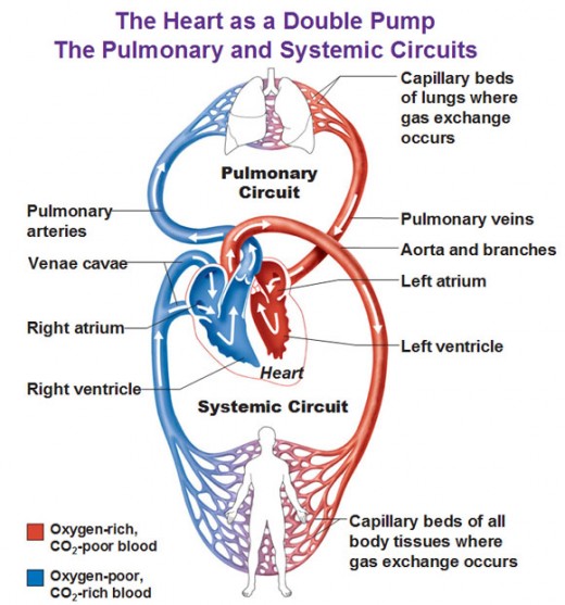
DIVISIONS OF CIRCULATION
The flow of blood is through two divisions of circulatory system, which are, systemic circulation and pulmonary circulation.
(1) Systemic circulation: this is also known as the greater circulation. The blood that is pumped from the left ventricle passes through a series of blood vessels of the arterial system and reaches the tissues. After exchange of materials at the capillary level, the blood enters the venous system and returns to the right atrium of the heart, and from the right atrium, it enters the right ventricle. Thus, through the systemic circulation oxygenated blood is supplied from the heart to the tissue and the deoxygenated blood returns back to the heart from the tissue.
(2) Pulmonary circulation: this is also called the lesser circulation. In this circulation, blood is pumped from the right ventricle to the lungs through the pulmonary arteries. Gaseous exchange occurs between the lungs and alveoli of the lung through the pulmonary capillary membrane. The oxygenated blood returns to the left atria through the pulmonary vein. Thus, the left side of the heart contains oxygenated blood or arterial blood while the right side of the heart contains deoxygenated or venous blood.


