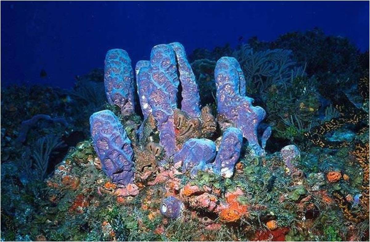
PHYLUM PORIFERA
PHYLUM PORIFERA
The sponges are the first metazoans (multicellular animals) that we will study. The principal features of phylum Porifera are listed below.
1. While some sponges are radially symmetrical, the majority of sponges are asymmetrical in body form. Sponges are considered to be at a cellular grade of construction; that is, they have cellular differentiation (tissues) without cellular coordination.
2. The outermost tissue layer of sponges is composed of cells called pinacocytes. In some sponges this outer tissue layer is syncytial while in others the pinacocytes are all distinctly separated from one another by cell membranes. The innermost tissue layer is composed of cells called choanocytes or collar cells (see S&S, p.45) which have flagella that beat to produce water currents through the sponge body. Between these two tissue layers is a gelatinous layer called the mesoglea (mesohyl). The mesoglea is not considered to be a tissue since it contains a number of different kinds of independently functioning cells. Each cell type in the mesoglea has a specific name, but the general term for all of these wandering cells is amoebocyte.
3. Some of the amoebocytes in the mesoglea are specialized for secreting a skeleton. The sponge skeleton may be composed of mineral spicules, spongin fibers or a combination of these two, depending on the kind of sponge. Spicules may be calcareous (composed of Ca CO3) or siliceous (composed of H2Si2O7). Spongin fibers are composed of a sulfur-containing schleroprotein.
4. Water enters the body of a sponge by way of a number of minute incurrent pores or ostia. Water leaves the body by way of one or more large excurrent pores or oscula. Within the body of the sponge, the water may pass through a large cavity (the spongocoel) through a system of canals and chambers, or through a combination of these two.
5. Movement of water through the sponge body is accomplished by the beating of the choanocyte flagella. The choanocyte cells line either a spongocoel or a number of small chambers, depending on the sponge. A choanocyte cell consists of a nucleus, one or more vacuoles, a long flagellum and a delicate, collarlike structure which surrounds the base of the flagellum. Electron microscope studies show the collar of a choanocyte to be composed of a circular arrangement of microvilli-like structures extending outward from the cell body. The rotary motion of the flagellum forces solid food particles in the incoming water to adhere to the outside surface of the collar. The streaming protoplasm of the collar transfers the food to the collar base where ingestion can occur.
6. Sponges may be divided into three basic grades or types based upon the arrangement of their water canal systems. Note that these grades or types are not taxonomic groupings. The three types of sponges are described below and are shown diagrammatically.
· Asconoid Type - Water entering the sponge passes through ostia which are actually openings within doughnut-shaped cells called porocytes, which are found only in asconoid sponges. The water enters the large central cavity called the spongocoel, which is lined with choanocytes. Water exits from the spongocoel through a single large osculum.
· Syconoid Type - Water enters the sponge through ostia which are openings between cells, rather than within cells as in asconoid sponges. Water then passes into radially arranged incurrent canals which lead to flagellated chambers lined with choanocytes. Water leaves the flagellated chambers by way of excurrent canals that lead to the spongocoel, which is lined a simple flat epithelium. Water exits from the spongocoel by way of a single large osculum. Note that the body wall of syconoid sponges is thicker than that of asconoid sponges and that the syconoid spongocoel is not lined by choanocytes as is the asconoid spongocoel.
· Leuconoid Type - The ostia of a leuconoid sponge are like those of a syconoid sponge. These ostia lead into a complex system of canals and flagellated chamgers that penetrate the very thick, dense mesoglea. There is no spongocoel in a leuconoid sponge. Rather, water reaches the oscula by way of large excurrent canals. The complex canal system of leuconoid allows for greater surface area over which water may pass and consequently creates an increased area for food and oxygen uptake and for waste removal. It is not surprising, therefore, that leuconoid sponges are the largest in size of all the types and that the vast majority of sponges are leuconoid.
7. Sponge taxonomy is based on skeletal composition. The four classes in phylun Porifera are listed below along with distinguishing characteristics for each class. The grades of sponges found in each class are given in parenthesis, although this is not distinguishing since there is overlap between the classes.
a) Class Calcarea - contains sponges having calcareous spicules with 1 to 4 rays. (asconoid, syconoid, leuconoid)
b) Class Hexactinellida - contains sponges having siliceous spicules with 6 rays. These spicules are often fused to form a beautiful lattice-like cylinder, as in the so-called Venus' flower basket. (syconoid)
c) Class Demospongiae - contains sponges having siliceous spicules (not 6-rayed) and/or spongin fibers. (leuconoid)
d) Class Sclerospongiae - contains sponges having an internal skeleton of siliceous spicules and spongin fibers plus an outer encasement of calcium carbonate. Only six species from the Caribbean have been described to date. (leuconoid)
8. Sponges are capable of both sexual and asexual reproduction and they also have great powers of tissue regeneration and reassociation. Sexual reproduction is accomplished by production of eggs and sperm which unite to form a zygote. The zygote divides repeatedly to produce a free-swimming larval form. Depending on the sponge, this larva may be either a uniformly ciliated parenchymula larva or an amphiblastula larva, which has flagella only at one pole (Fig. 2.7, p.55 of S&S). The larvae eventually settle and metamorphose into the adult form.
Sponges may reproduce asexually by budding. In addition, all freshwater sponges and some marine forms produce resistant overwintering bodies called gemmules. These gemmules consist of aggregations of food laden amoebocytes surrounded by a resistant covering. They are produced during periods of cold or drought and can survive to produce a new sponge body when conditions improve (Fig. 2.8, p.55 of S&S).
In today's laboratory you will examine the structure of the sponge species available and then perform experiments on the reassociation of porifera cells.
ASCONOID SPONGES
Asconoid sponges are the simplest and most primitive sponge architectural type and are all relatively small due to their inefficient filtering system. Asconoid structure is demonstrated in Leucosolenia. Obtain a small colony of Leucosolenia sponges and place it in a dish filled with seawater. Examine it under a dissecting microscope. Does it respond to a stimulus? Do you detect movement? To observe the filtering mechanism of Leucosolenia, prepare a dilute suspension of carmine powder and seawater and then gently place a drop of the suspension near the colony. Describe the water movement through Leucosolenia. Where do the carmine particles enter? Where do they exit? Do particles enter the sponge at the same velocity that they exit? Explain. Do you see budding on your Leucosolenia colony? If so, where are the buds positioned? Examine the colonial structure of Leucosolenia. Describe how you think the ultimate colonial form develops from a single sponge tube.
From your Leucosolenia colony remove a single sponge tube for closer examination and then rinse the colony in fresh seawater and return it to the holding tank. Cut the sponge tube longitudinally into two halves (from osculum to base) and place the two halves on a slide so that half the tube shows the inner surface and the other half shows the outer surface. Add a drop of saltwater and cover with a coverslip. Examine both surfaces under the compound microscope. Try to identify porocytes, pinacocytes, and choanocytes, or evidence of their presence (refer to S&S, pp. 45‑47 for diagrams). Next, tease apart the sections of sponge with a dissecting needle and examine for spicules and amoebocytes. Describe the shape and arrangement of the spicules. How are spicules formed? Do you see any evidence of this in your preparation?
Examine the prepared slides of asconoid sponges. The staining of these slides will make the cellular structures easier to identify.
SYCONOID SPONGES
Syconoid sponges are more complex than asconoid sponges. Syconoid sponges look like large asconoid sponges, having a tubular shape, and each individual has a single excurrent osculum. The body wall is thicker, however, and the spongocoel is lined with pinacocytes. The choanocytes line finger-like chambers (radial canals), which permeate the spongocoel (see pp. 50-51 in S&S). Because this arrangement provides a more efficient pumping system than the asconoid design, syconoid sponges are larger than asconoid sponges.
Obtain a single specimen of Scypha, a representative syconoid sponge. How does the colonial form of Scypha compare to Leucosolenia? Place your Scypha individual in a dish filled with seawater and examine it under the dissecting scope. Does it respond to mechanical stimulation? How does its filtration system compare to Leucosolenia? Section the Scypha and examine its body wall structure. Examine the mesoglea and characterize the spicules of Scypha (p.52 of S&S).
The prepared slides of a second syconoid sponge, Grantia, clearly shows wall structure, choanocytes, and spicules.
LEUCONOID SPONGES
Leuconoid sponges are by far the most complex architectural-type of sponge. Most leuconoids are colonial, and although individual oscula can be distinguished,it is difficult to separate individual members of the colony. The vast majority of sponges are leuconoid. Examine the external and internal anatomy of the leuconoid available in the laboratory (probably Microciona). How does itsfiltration system differ from the asconoid and syconoid sponges you examined? Examine the body wall structure (see S&S, p. 51). Characterize the spicules.
SPICULE COMPOSITION AND STRUCTURE
The systematics of sponges are based primarily on the composition and structure of spicules rather than on architectural plan. Spicules are composed of either calcium carbonate or silicon dioxide, and the skeleton may consist entirely of collagenous fibers (spongin) or a combination of spicules and spongin. See the introduction of this exercise (#8) for the characteristics of the four classes of Porifera. The class membership of sponges is easily determined using the following tests:
1. The organic matrix of sponges (spongin) dissolves when boiled in 5% sodium hypochlorite solution. Place small pieces of sponge tissue in 1-2 ml of the sodium hypochlorite solution in a testtube to carry out this test. Boil the mixture for a couple of minutes by placing the test tube in a beaker of boiling water. After cooling, examine under the compound microscope. Are spicules present? This technique is also used to remove spongin from spicules to examine spicule form.
2. The inorganic chemical nature of spicules is determined by drawing acetic acid or dilute hydrochloric acid under the cover slip of a wet mount of spicules. Calcium carbonate spicules dissolve when treated in this manner, while silicon dioxide spicules are not influenced by the treatment.
3. Examination of the shape of spicules found in a sponge are also important taxonomic characteristics (see S&S, p. 52, for representative spicule morphology).
v Determine the class membership of the sponges available in the laboratory.
REASSOCIATION
Sponges have remarkable powers of regeneration. A complete sponge can regenerate from only a handful of cells. In the natural environment this means that when a sponge is disturbed by a predator or physical disturbance, remaining fragments are able to form new individuals and colonies. The impressive regeneration ability of sponges is due to the loose organization of cells in individuals. Individual cells and unorganized clusters of cells are able to reassociate and organize into new individuals. In today's laboratory we will examine the reassociation phenomenon of sponges.
vProcedure: Obtain a few milliliters of the suspension of Microciona cells available in the laboratory. This suspension was prepared by pressing pieces of fresh Microciona through a silk bolting cloth into seawater. Examine a drop of the suspension under a compound microscope. It should consist of small clumps of cells.
Prepare a series of dilutions in seawater from the original suspension so that you have samples of decreasing densities. Make your dilutions 100%, 50%, 25%, and 10% solutions of the original suspension. Fill 2 small fingerbowls two-thirds full of cool seawater; place a Syracuse watch glass on the bottom of each fingerbowl and put two slides on each watch glass. Before placing the slides on the watch glasses, individually number each slide. Using a Pasteur pipet, gently dispense equal aliquots ( 2 drops) of each suspension onto the slides. Place two treatments (dilutions) on each slide and have all four dilutions represented in each fingerbowl. Be very careful not to allow mixing of the dilutions on the slides. Be sure you record the positions of the different dilutions on each slide. Clearly label your fingerbowls and have one stand at 4°C and the other at room temperature for 24 hours. After 24 hours, gently remove the slides, cover with a coverslip, and examine under a microscope. Systematically count the number of aggregates in each dilution and compare the size distribution of aggregates in each dilution. Record all your data and observations. How do cell density and temperature influence aggregation? What form do the aggregates take? How are different cell types arranged in the aggregates?
Place a fresh drop of the Microciona suspension on a slide and examine it during the course of the laboratory under l00X. Do you see any evidence of aggregation?
Following the procedures outlined above for examining reassociation in Microciona, prepare similar solutions of a suspension of the cells of two sponge species (Microciona & Haliclona). Prepare a fingerbowl incubating the four interspecific dilutions and let it stand for 24 hours at 15°C. Compare these results with the Microciona suspension. What has happened to the cells of the two species? How might segregation of the two cell types have occurred? What other experiments could be performed to test this phenomenon further?
scyphaScypha, also called sycon, genus of marine sponges of the class Calcarea (calcareous sponges), characterized by a fingerlike body shape known as the syconoid type of structure. In the syconoid sponges, each “finger,” known as a radial canal, is perforated by many tiny pores through which water passes into a single central cavity. The water exits through an oscule, or larger opening, at the tip. Water is driven through the sponge by the beating of many hairlike cilia lining the central cavity. Scypha species grow to only about 2 or 3 cm (about 1 inch) in length.
The Body of a Sponge
Sponges (phylum Porifera) are the simplest animals. For example, Scypha, a small marine sponge measuring only a few centimeters, resembles a simple tube with an opening at one end (Figure 23-4). Unlike most animals, sponges lack true tissues and organs. Most of the different types of cells in a sponge are relatively unspecialized
The body of most sponges consists of two layers of cells separated by a jelly-like material. The outer layer of cells protects the interior of the sponge and also has many pores (holes) through which water can enter the sponge. The inner layer of cells lines the central cavity of the sponge. These cells, called collar cells, have flagella. Wandering through the jelly-like material are cells called amoebocytes that pick up food from the collar cells, digest it, and carry the nutrients to other cells. Amoebocytes also transport oxygen, dispose of wastes, and can change into other cell types, such as support structures. In some sponges these support structures are rigid. In other types of sponges the structures are composed of a flexible protein called spongin. It is these flexible sponges that are harvested for use as bath sponges.
Sponges ingest food by the action of their collar cells. The flagella of collar cells generate water currents that move water through the sponge's pores and into the central cavity. As water flows through the sponge's body, it is filtered for food particles (mostly bacteria) in the water. Collar cells trap the food particles in mucus. Amoebocytes then engulf the particles and transport them to other cells. The water drawn in through the pores exits through the large opening at one end of the sponge.
Sponges live singly or in clusters formed by budding. Budding is a form of asexual reproduction in which new sponges develop from an outgrowth of the parent organism. Most species of sponges have great powers of regeneration. Small fragments of a sponge body can grow into an entire new sponge. However, sponges can also reproduce sexually. Most sponges have both male and female gamete-producing structures in the same organism. The union of egg and sperm cells form zygotes that develop into flagellated larvae.
Adult sponges are sessile, meaning they are anchored in place. They can't run and hide. Sponges have chemical defenses that protect them from possible predators, disease organisms, and parasites. These defenses include toxins that keep predators from eating the sponges and powerful antibiotics that fight bacterial infections. Researchers are studying these unique sponge chemicals for new drugs to fight human disease.
Although sponges are animals, they have several protist-like features. Their relatively simple structure supports evidence from DNA analysis that sponges evolved very early from the protists that were ancestral to the animal kingdom. The modern protists that are most closely related to animals are colonies of protist cells that are very similar to the collar cells of sponges. Later in the chapter, you will learn about a hypothesis for how such colonial protists may have given rise to the animal kingdom.
Diversity of Sponges
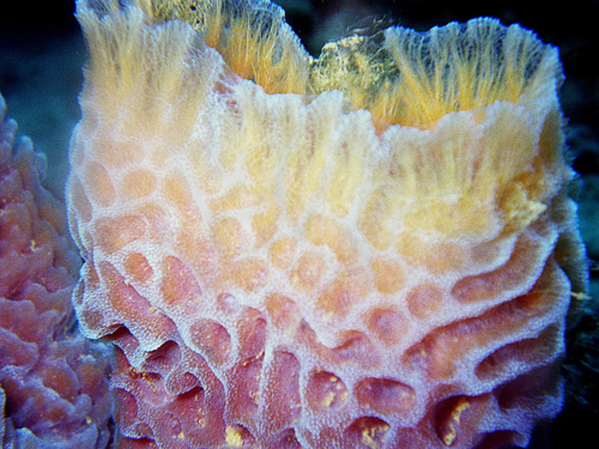
The 9,000 known species of sponges are diverse in shape, size, and color. Some sponges consist of a single cylinder, while other sponges branch out irregularly over the seafloor or lake bottoms. Some sponges, like Scypha, are quite small. Others can reach heights of 2 meters.
