13 Year-Old Back to School One Week After Rare Brain Cyst Surgery

Amazingly Quick Recovery
First Came the Vomiting
In January 2006, 13 year-old Jimmy Graves of Crystal River, Florida was having intermittent episodes of morning vomiting, shortly followed by excruciating headaches, unrelieved by medicine. He would wake up, eat two bites of cereal, and then go straight to the bathroom vomiting bile. This ritual became an unfortunate way of life for the Central Florida seventh grader. The vomiting would come at unpredictable times: in math class, after school, before bed, and other non-patterned times.
Per the recommendation of a local walk-in clinic, Jimmy was referred to Nemours Children’s Hospital in Orlando for an appointment with a pediatric gastroenterologist. Suspecting GERD (Gastroesophageal reflux disease), the doctors at Nemours scheduled Jimmy for an endoscopy. They inserted a little chip at the bottom of his esophogus, along with a portable transmitter to read the acid levels from Jimmy's stomach. After several days of readings, the report came back normal. Jimmy did not have GERD. The doctors were perplexed by Jimmy's frequent episodes of vomiting. They recommended he keep a food diary to correlate any relationship between certain foods and the unexplained vomiting.
Next Came the Headaches...
Over the next several months, Jimmy continued to wake up with vomiting of bile and painful headaches, not daily but intermittently. On one such morning, he took 2 Rapid Release Tylenol. He still went to school, but within an hour and a half, Jimmy was sent home by the school nurse, still suffering from a severe headache. Jimmy continued to have headaches every week.
After catching a cold, Jimmy visited his pediatrician. Upon mentioning the headaches to his doctor, Dr. Dacelin St. Martin, he asked Jimmy a series of questions to rule out migraines. Based on Jimmy's responses, the doctor decided that it didn't fit the migraine pattern, so he ordered an MRI of Jimmy’s brain.
Jimmy was sent to a local imaging center for an MRI, and they put him in the open MRI scanner. The open MRI showed a "signifcant dilation the posterior horn of the right lateral ventricle". The radiologist suggested another MRI be prescribed for a closer look at his brain on the enclosed scanner. After going in for a closer look, the radiologist found a large intraventricular cyst in the Choroid Plexus region of the right lateral ventricle (on the right side of Jimmy's brain).
MRI Image of Jimmy's Brain

The "Conservative" Approach... Suffering
Jimmy was referred to a pediatric neurologist in Tampa, Florida. The neurologist suggested Jimmy's headaches were migraines, unrelated to the cyst, and the cyst was an incidental finding. She prescribed Topamax for the headaches, and referred Jimmy for an appointment with a pediatric neurosurgeon.
Jimmy next saw a pediatric neurosurgeon in St. Petersburg, Florida. She said that the cyst was unrelated to the headaches, and she believed in a conservative approach - "watch the cyst and wait", even though Jimmy was clearly suffering. She recommended that Jimmy have a second opinion. From a handful of pediatric Neurosurgeons in the state of Florida, Dr. John Ragheb at Miami Children’s Hospital came highly recommended.
The True Diagnosis and Prognosis
On Tuesday, August 1, 2006, Jimmy saw pediatric Neurosurgeon, John Ragheb, M.D. He was the Director of the Neurosurgery Department at Miami Children’s Hospital. Dr. Ragheb listened carefully, and pieced together Jimmy’s history.
Dr. Ragheb said that Jimmy’s cyst was congenital (he was born with it). He made the correlation between the morning headaches and vomiting; a classic result of increased intracranial pressure. The cyst was partially blocking the flow of CSF in the brain ventricle, and was blocking it more in the morning upon wakening. Dr. Ragheb said that Jimmy’s cyst location was rare. The size of the cyst was enough to warrant brain surgery to prevent further growth, and exaggeration of the symptoms causing the suffering.
Rare Congenital Brain Cyst Causing Headaches and Vomiting

Nicklaus Children’s Hospital (formerly known as Miami Children's Hospital)

New Frontier of Brain Surgery
The treatment for Jimmy's brain lesion would be endoscopic cyst fenestration, with Brain Lab Navigation. The doctor makes a small hole in the skull near the cyst, and puts a tiny scope with a camera in the brain. He takes a biopsy of the cyst and its contents. If it is simple CSF fluid in the cyst, then the cyst will be drained.
Jimmy was scheduled for endoscopic cyst fenestration surgery at Miami Children's Hospital on Monday, October 30, 2006.
Brain Lab Navigation Surgery... As Seen on Grey's Anatomy
Jimmy endured Neuroendoscopic cyst fenestration surgery with Brain Lab Navigation. The surgeon drilled a small, 3 cm hole into the skull. The cyst was extremely large (he compared the size of the 3cm hole to the size of the cyst, and said it was like an elephant to a mouse). The contents of the cyst were drained and consisted of clear spinal fluid.
After undergoing brain surgery on Monday, Jimmy was discharged from Miami Children's Hospital on Wednesday. He went home, and returned to his eighth grade gifted classes at Crystal River Middle School 5 days later.
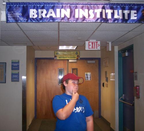
Images from Hospital Stay


Happily Ever After...
The tidy, 2 inch surgical scar hiding under Jimmy's Boston Red Sox hat is the only visible evidence that this now healthy high school senior had brain surgery four years ago. Jimmy's headaches and vomitting have completely disappeared. Dr. John Ragheb at Miami Children’s Hospital made a big difference in the quality of Jimmy's life, and the life of his family. I know personally, because Jimmy Graves is my son.
Jimmy Graves and me

Helpful Resources
- What are arachnoid cysts?
Arachnoid cysts are fluid filled sacs or cavities that appear on the arachnoid membrane and can present in many locations on the brain and/or spinal cord. Majority of these cysts are discovered incidentally and do not merit surgical intervention but - Dr. John Ragheb, MD - | Nicklaus Children's Hospital
Bio for Neurosurgeon John Ragheb, M.D. - Ventriculoscopic surgery for arachnoid cysts in the lateral ventricle: a comparative study of 21 con
Ventriculoscopic surgery for arachnoid cysts in the lateral ventricle: a comparative study of 21 consecutive cases. - Dacelin St. Martin, M.D.
Bio of Pediatrician Dacelin St. Martin, M.D.


