Acute Rheumatic Fever: Health Significance, Epidemiology, Course And Progression Of The Infectious Disease
Medical Care On Acute Rheumatic Fever

Acute Rheumatic Fever
Rheumatic fever is not an uncommon disorder seen in the pediatric age group worldwide. It is clinically characterized by fever, polyarthritis, carditis, chorea, skin manifestations and pleurisy. The underlying cause is hypersensitivity reaction to group A streptococci. Rheumatic fever follows an attack of streptococcal pharyngitis after 2 to 3 weeks. Rheumatic valvular disease accounts for a major proportion of cardiac morbidity occurring below 40 years of age in India.
Epidemiology: Though, rheumatic fever is worldwide in distribution, it is more common in the developing countries. A Group A hemolytic streptococci may occur as commensals in the throat in a small proportion of people. Sometimes, these streptococci become virulent and produce outbreaks of upper respiratory tract infections, especially in children living in closed communities like schools and dormitories. Since overcrowding and poor living conditions predispose the spread of streptococcal infection, rheumatic fever is more common in the poorer, socio- economic groups. The risk of streptococcal tonsillitis is 0.3 to 3%. The most vulnerable age group is 5 to 15 years, though any age may be affected. In West African, the mean age of affection is 5.5 years. Both sexes are affected and a familial predisposition is noticed in many cases.
Pathogenesis
In 80% of cases, the disease follows streptococcal tonsillitis. In 20 to 30% of cases, the episode of tonsillitis may go unnoticed. Over 80% of subjects with rheumatic fever show rise in antistreptococcal antibodies within two months. The antigen contained in streptococcus show similarity to several tissue antigens in the human body, especially myocardial sarcolemma. Antibodies produced against streptococcal antigens cross react with tissue antigens resulting in a type II antigen antibody reaction and consequent inflammation. Lesions are most evident in the connective tissue.
Pathology: The pathological process consists of an exudative stage which is seen in the acute phase and a proliferative stage which is a more prolonged process. In the exudative stage, fibrinoid necrosis of the connective tissue is seen. There is edema of the collagen fibers which later undergo fragmentation.
The hallmark of the proliferative phase is the formation of Aschoff bodies. These are small granulomas consisting of a central zone of fibrinoid necrosis surrounded by round cells, epithelioid cells, Aschoff giant cells and fibroblasts. This histological appearance is similar in all tissues showing rheumatic lesions.
Heart: All the layers are involved to produce endocarditis, myocarditis and pericarditis (pancarditis). Endocardidits affects the valvular or mural endocardium. When the valvular or mural endocardium heals, it results in scarring and deformity of the valves. Scarring after mural endocarditis may be seen as the MacCallum’s patch in the posterior wall of the left atrium. Affection of the myocardium may be mild or severe. Lesion in the pericardium is a fibrinous inflammation. The pericardium shows a ‘bread and butter’ appearance macroscopically. Pericardial effusion may develop. Adhesive pericarditis may follow rarely.
Joints: Rheumatic lesions involve large synovial joints producing acute synovitis. Effusions develop which clear up completely without any residual deformity.
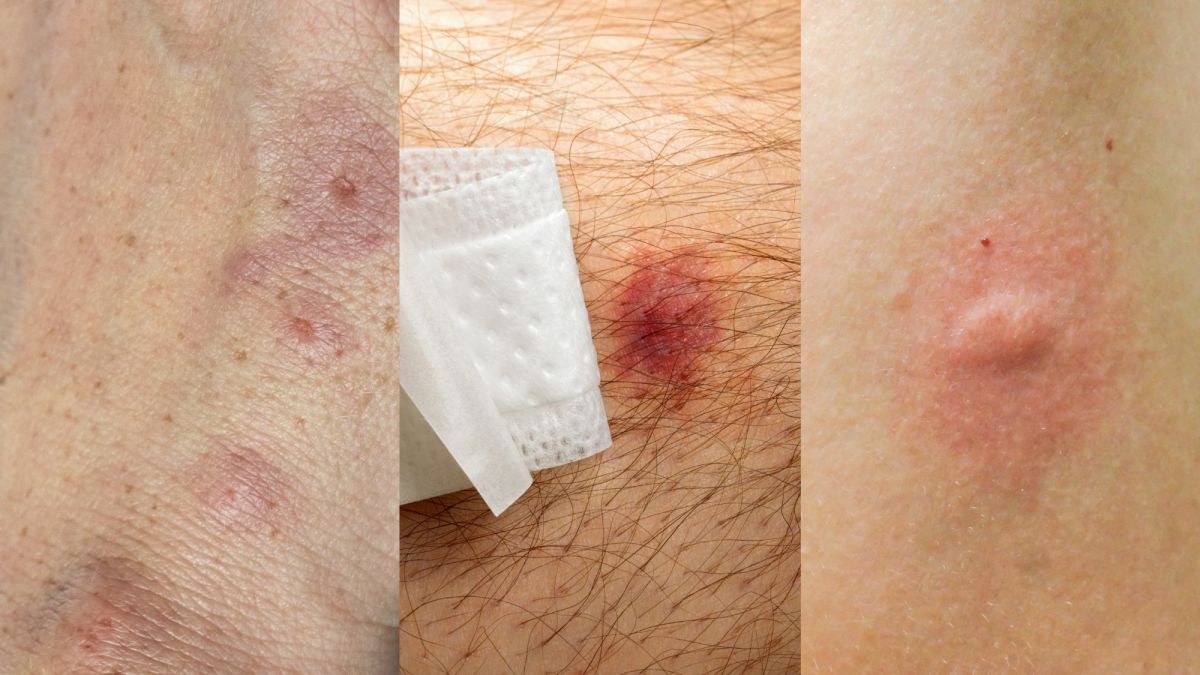
Other Organs: Rheumatic granulomas found in the subcutaneous tissues, are seen as subcutaneous nodules. Respiratory involvement manifests as a pneumonia with or without evidence of pleurisy. The basal ganglia of the brain are affected. The lesions take the form of aseptic focal encephalitis which clears completely.
An Acute Rheumatic Fever Patient

Course And Disease Progression
Rheumatic fever tends to subside spontaneously over a period of weeks or months. The arthritis subside but the cardiac lesion progresses. Recurrence of streptococcal infection results in exacerbation of the rheumatic process and hence relapses invariably occur. The course of the disease extends over several months or even a few years with remission and relapses. The risk of developing fresh cardiac lesions and worsening of the existing lesions is increased with successive relapses. Mortality in the acute phase is due to sudden cardiac failure or heart block.
© 2014 Funom Theophilus Makama


