African Trypanosomiasis: Health Implications, Transmission And Its Pathology
The African Trypanosomiasis Infection

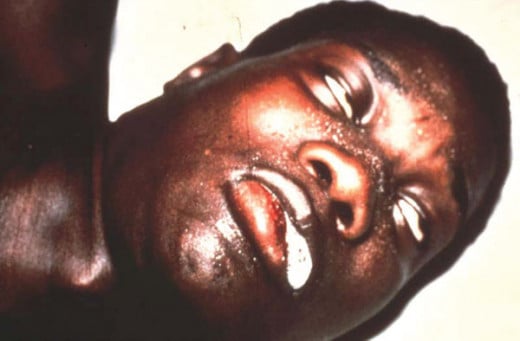
A General Overview Of Sleeping Sickness
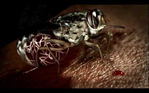
African trypanosomiasis or sleeping sickness is caused by Trypanosoma gambiense or T. rhodesiense. These hemoprotozoa are transmitted by the bite of infected tse tese flies (Glossina).
Geographical distribution: Trypanosoma gambiense is found in West Africa, Congo, Southern Sudan and Uganda. Trypanosoma rhodiense is found in Central and Eastern Africa. Endemicity is closely related to the distribution of the vector tsetse flies and the reservoir hosts.
Trypanosomes are hemoflagellates seen as actively motile spindle shaped organisms. In leishman stained preparations, trypanosomes measure 14 to 33 X 1.5 to 3.5 microns. They possess a central nucleaus and a Kinetoplast situated near the posterior end. A flagellum arises from the blepharoplast situated close to the kinetoplast and passes forwards along the free margin of the undulating membrane and becomes a free flagellum protruding membrane and becomes a free flagellum protruding forward from the anterior end. Trypanosomes are seen actively moving in the plasma in fresh preparations. Trypanosoma gamniense and Trypanosoma rhodesiense are morphologically similar, but the latter is more virulent in laboratory animals.
The Tse tse Fly Causing The Sleeping Sickness

Infectious Diseases
Transmission And Pathology Of Sleeping Sickness
Transmission
The vectors are mainly Glossina palpulis (G. fuscipes) and G. tachinoides for Trypanosoma gambiense and G. morsitans, G. Pallidipes and G. swynnertoni for Trypanosoma rhodensiense. Both sexes of tsetse flies suck blood and transmit the disease. They live for 2 to 5 months.
Trypanosoma rhodiense is a widespread parasite of several herbivorous animals in endemic areas and these form the reservoir. Man is secondarily infected from them through the vectors. Man to man spread is also possible. Tsetse flies get infected by feeding on infected animals or man. The parasites multiply in the gut of the fly and pass anteriorly to the proventriculus and the salivary glands. The infective metacyclic forms develop and multiply there further and pass into the proboscis after 3 to 7 weeks. The fly remains infective for its life. The metacyclic forms are introduced into the host during the next bite. The metacyclic forms transform into trypanosomes within 48 hours and multiply in the tissue space. Ultimately, they pass to lymph nodes and the blood stream and then reach several organs.
Pathology
Lymph nodes and the central nervous system (CNS) show macimal lesions. There is localized or generalized lymph node enlargement. The nodes are congested and show hemorrhagic areas which contain trypanosomes. Later, these undergo fibrosis. Liver and spleen are enlarged.
Affected organs show infiltration by lymphoid and reticuloendothelial cells. In the CNS, the main lesion is leptomeningitis seen over the brain and spinal cord. Histologically, there is perivascular cuffing with lymphocytes and plasma cells. Later, trypanosomes invade the brain tissue, particularly the frontal lobe, pons and medulla. Parasites are seen in the cerebrospinal fluid during the meningitis stage. Other changes include rise in pressure, moderate rise in proteins and lymphocytosis. Complement-mediated hemolytic anemia may develop in some. IgM class antibodies are demonstrable in the serum and CSF.
© 2014 Funom Theophilus Makama


