American Trypanosomiasis: Health Implications, pathology, Clinical Manifestations, Diagnosis, Treatment And Control

Chagas Disease In Manifestation

The General Clinical Overview Of Chagas Disease
This disease is caused by Trypanosoma Cruzi (Schizotrypanum) which is transmitted by certain reduvid bugs to man. It is seen in all countries of central and South America.
Trypanosoma cruzi is a broad trypanosome about 20u in length and is C-shaped. The motile stage of the parasite is found only for a short period after infection. It soon develops into the cells of mesenchymal origin. Reduvid bugs of the genera Triatoma, Rhodinus and Eratyrus are the principal vectors. Reservoir hosts are formed by the dogs, cats, Opposums and armadillos. Reduvid bugs are infected by taking blood meal and in 6 to 15 days, infective metacyclic forms are passed in feces. Organisms enter the body when the feces is smeared on to the wound.
Pathology: At the site of inoculation, the organisms multiply and produce a local lesion- chagoma. They are seen in the blood stream for a short period. They are taken up by macrophages later. Trypanosoma Cruzi multiplies and produces focal lesions in the cardiac and skeletal muscles and neuroglia. Cardiomyopathy develops as a result of cardiac involvement. Granulomatous lesions occur in the spleen, liver and lymph nodes. CNS involvement in children leads to meningo-encephalomyelitis.
A Bite From The Tsetse Fly In Chagas Disease
Infectious Diseases

Clinical Manifestations Of Chagas Disease
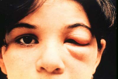
In children, chagas disease occurs in an acute form, presenting with Chagoma, generalized lymphadenopathy, fever, myocarditis and rarely meningoencephalitis. The initial lesion presenting with unilateral edema of the conjunctiva and adjacent tissues is called Romana’s sign. In the chronic type, cardiomyopathy and in some cases dilatation of the esophagus, colon and the small gut are seen. The alimentary lesions are due to destruction of the Auerbacj’s plexus.
Diagnosis: It is established by the demonstration of trypanosomes in the peripheral blood or its leishmanial form by muscle biopsy and by serological tests. Xeno-diagnosis is established by feeding the uninfected vectors on suspected cases and examining the hindgut two weeks later for the presence of the parasite. Serological tests like complement fixation test, indirect fluorescent antibody test and hemagglutinin test are helpful.
Treatment: Primaquine has been found useful in a dose of 15 mg daily for about 6 months in acute Chagas disease. At present, Nifurtimox (Lampit) a nitrofurantoin, is available for oral use. The dosage is 8 to 10 mg?Kg/day for adults and 15 to 20 mg?Kg/day for children and the drug has to be administered for 120 days. Cure rates of 80% in acute and 90% in Chronic cases have been claimed. The drug is toxic and hence has to be given under supervision. Benzimidazole (Rochagan) in an oral dose of 6 mg?Kg for 30 to 60 days is also effective. This has to be given under supervision.
Control: Improvement in housing and sanitation helps to limit the vectors. Gammaxane spray is useful in eliminating the reduvid bugs.
© 2014 Funom Theophilus Makama
")


