Etiology, Diagnosis, Clinical Presentations, Investigations And Treatment In Hodgkin’s Lymphoma

Diagnosing Hodgkin Lymphoma
A Quick Overview
These are malignant tumours arising from lymphoid tissue. Lymphomas remain confined to lymph nodes and other lymphoid structures in their early stages. Lymphomatous cells in varying numbers are seen later in the bone marrow and the peripheral blood. Lymphomas may start unicentrically or multicentrically. They have been classified broadly into Hodgkin’s Lymphoma (HL) and non-Hodgkin’s lymphoma (NHL). Hodgkin’s lymphoma is characterized by the presence of Sternberg-Reed cells which are absent from the latter. The course, prognosis and response to treatment are different in the two types. The nature of the abnormal cell in HL is not very clear. Non Hodgkin’s lymphoma is known to be a neoplasm arising predominantly from B lymphocytes.
Etiology
As in the case of leukemias, several possibilities have been postulated. These include genetic predisposition, viral infections, immunological factors long-term toxicity to drugs like cyclophosphamide and exposure to irradiation and chemicals. There are may reports of lymphomas occurring in clusters in closed ethnic groups, and showing a seasonal prevalence. These phenomena suggest a common environmental factor.
Epidemiology
Lymphomas form 1.9% of all cancers in many general hospitals in Nigeria. Among the lymphomas, 35- 43% is HL and the rest is NHL.
Lymphatic malignancy

Pathology
Pathology: Hidgkin’s lymphoma generally arises unicentrically and spreads to other lymphatic tissues by contiguity and later to non-lymphatic organs as well. The diagnostic Sternberg-Reed (SR) cell is a large multinucleated cell with mirror image nuclei and it is though to be derived from histocytes. The lymph node architecture is destroyed and replaced for abnormal tissue. Four histological patterns have been recognized.
Rye (1965) classification
- Lymphocyte predominant (10%): There is predominance of lymphocytes with a few SR cells. Nodular and diffuse patterns are recognizable. This has the best prognosis.
- Nodular sclerosis (30-40%): In this form, the tumour is arranged as nodules which are encircled by collagenous bands extending from the capsule. A variant of the SR cells- the lacunar cell- may be present. Mediastinal lymph node enlargement is more common.
- Mixed cellularity (20-30%): There are numerous SR cells and moderate number of lymphocytes.
- Lymphocyte depleted (10-20%): The pattern may be reticulate or diffuse fibrosis. In the former, there is dominance of SR cells and depletion of lymphocytes. In the latter, the lymph node is replaced by disorderly connective tissue containing a few lymphocytes and a few SR cells.
The nodular sclerosis type maintains its distinct histology throughout whereas the other types may progress to one or the other or show overlap when followed up. In India, mixed cellular type is more frequent than nodular sclerosis. The prognosis becomes progressively worse as one moves from the lymphocyte predominant type to the lymphocyte depleted types.
Hodgkin’s lymphoma may be encountered at any age, but it is more common in the second decade. Males are affected twice as frequently as females. The majority of patients come in an advanced stage, the disease having been present for more than six months.
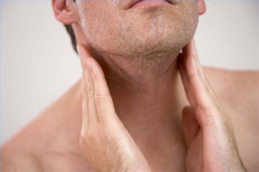
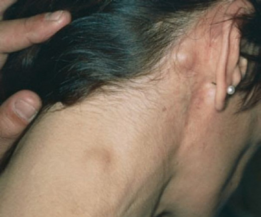
The most frequent symptoms is painless asymmetrical circumscribed enlargement of the lymph nodes, which are rubbery in consistency. In over 75% cases, cervical groups are affected. Axillary, mediastinal and inguinal groups are involved less frequently. Involvement of the retroperitoneal lymph nodes is not rare. In the absence of CT scanning or lymphangiography, these can be demonstrated only at laparotomy. The lymph nodes reach moderate to large sizes. They do not soften, break down or ulcerate.
Moderate splenomegaly occurs in over 50% of cases and hepatomegaly in a smaller number. These organs are firm and non-tender. Hepatocellular or obstructive jaundice may occur.
Constitutional symptoms occur in the majority of cases in the later stages. Pel- Ebstein fever consists of a few days of high swinging pyrexia followed by an afebrile period. Fever is commonly due to concomitant infection, but less commonly it is due to the malignant process itself. The patient rapidly loses weight and develops toxemia. Sweating, intractable pruritis and anemia develop in many cases.
Alcohol induced tenderness over the lymph nodes and bone is described as a diagnostic feature, but in Indian subjects, it is rate. Horner’s syndrome may occur due to pressure on the cervical sympathetic. Spinal root and cord compression and development of complications. Opportunistic infections such as herpes zoster, M. tuberculosis, cytomegalovirus, cryptococci and Candida spp, are common.
Treatment Of Hodgkin's Disease

Investigations And Diagnosis
Investigations
There may be normocytic normochromic anemia. In the advanced stage when the bone marrow is infiltrated, myelophthisic anemia develops. Mild eosinophilia may occur frewuently. In the advanced stage, lymphopenia develops.
Bone marrow involvement is seen in 10- 20% of cases and this is best demonstrated by trephine biopsy. As the disease progresses the immunologically competent T lymphocytes progressively diminish and this can be demonstrated in tuberculin test. With achievement of remission, the cell-mediated immunity recovers. Humoral immunity may be normal.
Bone involvement is accompanied by hypercalcemia, hyperphosphatemia and raised alkaline phosphatase levels. Hyperuricemia may develop. Hepatic alkaline phosphatase is raised. Serum copper is elevated during the active stages and it falls during remission.
Diagnosis
All cases presenting with chronic local or general lymphadenopathy in which a distinct cause cannot be established should be subjected to lymph node biopsy without delay. When several groups of nodes are affected, an entire node which is moderately enlarged should be selected for biopsy.
Differential diagnosis: All other causes of lymph node enlargement such as leukemias, tuberculosis, syphilis, filariasis, secondary carcinomatosis, nonspecific lymphadenopathy, toxoplasmosis, sarcoidosis and pseudolymphoma have to be excluded.
Drugs like streptomycin and phenytoin produce a type of immunoblastic lymphadenopathy (Pseudolymphoma), clinically resembling lymphoma. Withdrawal of the drugs results in resolution of the node.
© 2014 Funom Theophilus Makama
 or Premature Ovarian Failure (Pof)")


