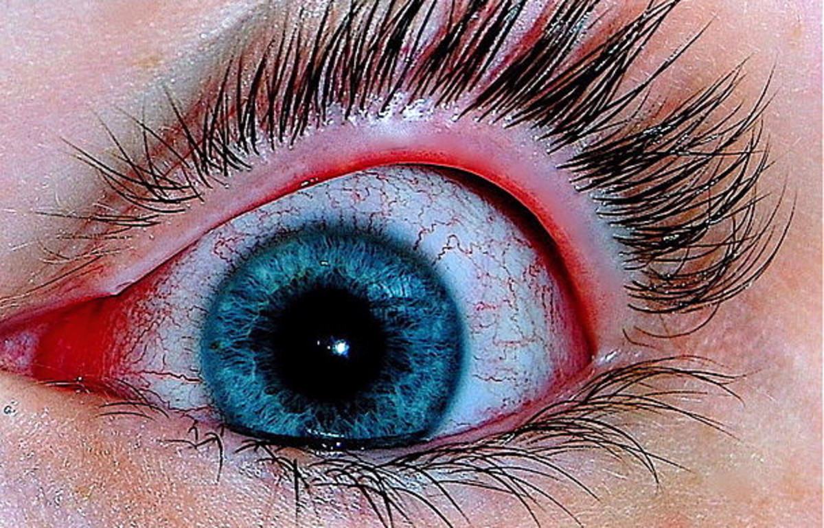
My Eye Floaters
Before I knew about eye floaters, I was always fascinated by the small worm-like figures in my eyes. I noticed them only occasionally and I had to focus hard to notice and track them as they moved about as the eyes moved. Then, one night at 55 years of age, I noticed a small lightning bolt appearing at the upper right corner of my right eye as I moved my eyes right to left. I did not see the small lightning bolt if my eyes remained stationary. It lasted for about two hours before disappearing completely.
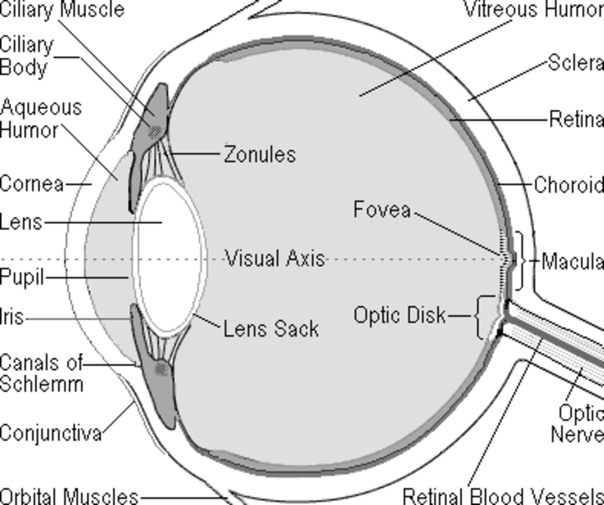
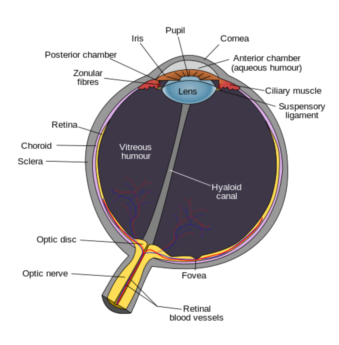
The next day, I noticed a drastic increase in the worm-like figures in my right eye. Some of them were quite large and began to interfere with my vision especially when I started to pay attention to them. I was a little bit worried so I looked up the condition on the Internet about Eye Anatomy. It was then I got to know what they were called, where they came from, and how they were developed.
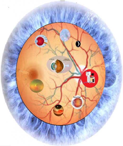
Research
From the Internet, I learned that the appearance of many floaters after the lightning bolt (or flash) could be the result of a retina tear that could result in blindness. So, I immediately checked into my HMO's emergency room. The doctor there also recognized the severe nature of the matter and arranged an appointment with the Ophthalmologist.
Diagnose
The Ophthalmologist first applied an eye solution to dilate both of my eyes. After the pupils were wide open, the Ophthalmologist used a light source and wore a magnifying glass to look into the back of my eyes in a very thorough and elaborate manner. After about 40 minutes of examination, the Ophthalmologist offered that there was no retina tear or detachment. I was greatly relieved to hear that. Afterward, I had to wear a dark-colored eyepiece for several hours to keep out the bright sunlight till the dilated pupils went back to normal.
Cause
The Ophthalmologist explained that my condition occurred when the vitreous gel, the thick fluid that filled the center of the eye, shrank and separated from the retina. This condition is called posterior vitreous detachment (PVD) which is often harmless. Sometimes, though, PVD can cause a tear in the retina.
At points where the vitreous gel is strongly attached to the retina, it can pull so hard on the retina that it can cause a tear in the retina. The tear allows fluid to collect under the retina and may cause the retina to detach and initiate the onset of blindness.
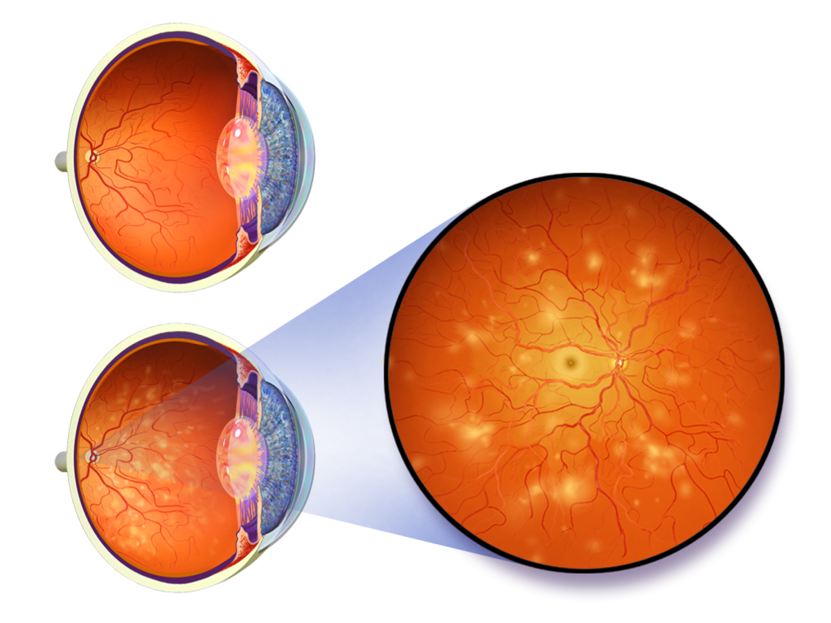
As the vitreous pulls on the retina, the photoreceptors are mechanically stimulated. Since the retinal cells are insensitive to pain, pressure, or temperature, the only stimulus that the retina responds to is 'light', thus, lightning bolt or flash. The tissues are torn from the area adjacent to the optic nerve head and become floaters.
Posterior Vitreous Detachment
PVD occurs in less than 10% of the people under 50 years of age but in more than 60% of the people who are over 70 years old. It is more common for people who are nearsighted or who have had an eye injury or have undergone eye surgery or who have had inflammation inside the eye. In my case, I am wearing a prescription eyeglass with a bad vision of around 20/1000. The Ophthalmologist also informed me that my left eye would undergo a similar process of the shrinking of the vitreous gel.
Aftermath
About two years later, my left eye went through similar conditions – flash then lots of floaters. I went to see the same Ophthalmologist with the same examination and the result – no retina tear. The Ophthalmologist said that there was no cure for the floaters but my vision should be fine as I got used to the floaters.
I needed to see the Ophthalmologist only when I noticed the occurrence of flashes or more floaters. I am now 70 years old and I have not experienced any more abnormal eye conditions except for worsening nearsightedness.
I have learned to tolerate the floaters which are more noticeable in the daytime and have not diminished in their quantities. I still go to see the optometrist for my glass prescription. I find that the Retina Optomap Exam machine is very useful. It takes a scan of the retina in less than a second without pupil dilation. The image taken can be viewed on a computer screen to check for the overall health of the eye.
Pyogenic Granuloma
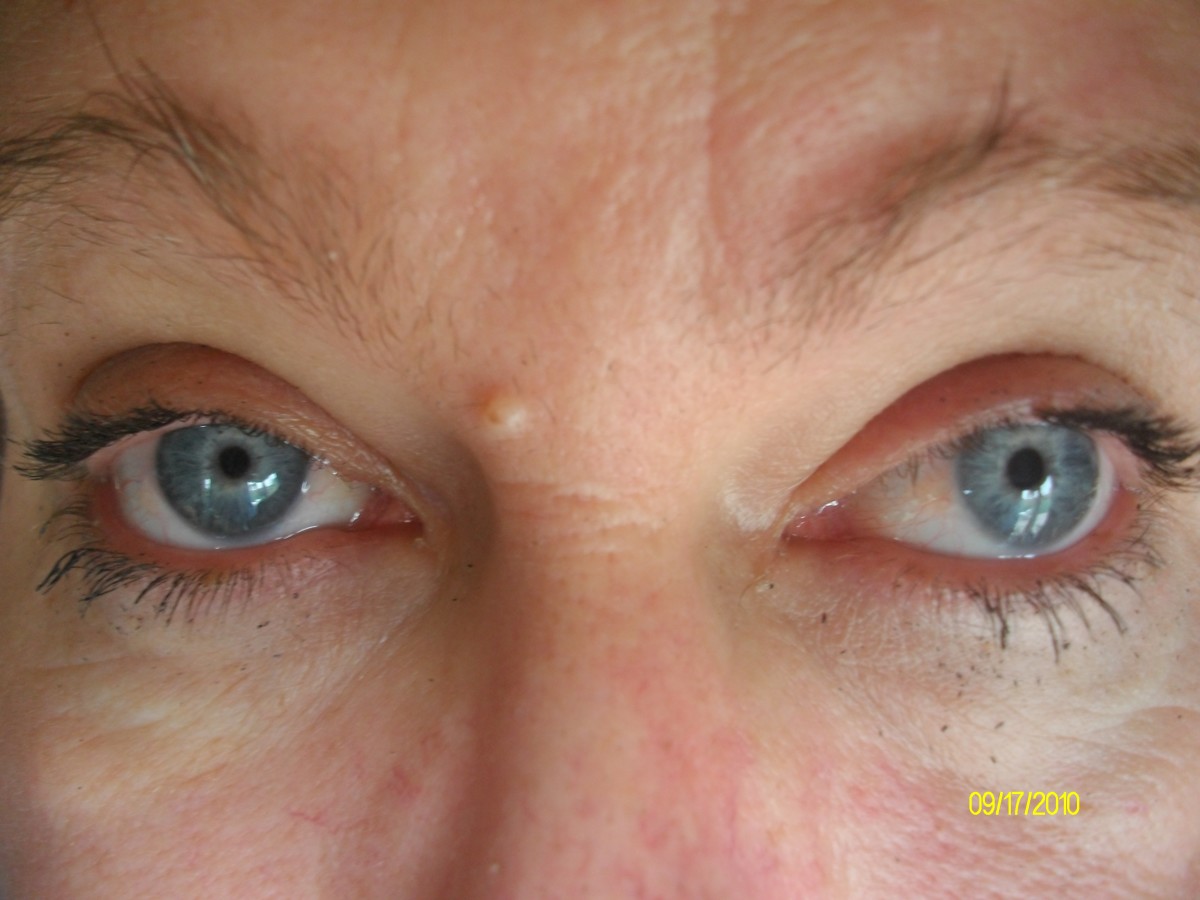
I also noticed a small growth inside my lower right eyelid. It had been there for at least 40 years. Since it never bothered my vision, caused no discomfort, or grew any bigger, I did not pay much attention to it. Recently, for curiosity, I looked it up on the Internet and found what it was called, where it came from, and how dangerous it could be.
It is called Pyogenic granuloma which is a relatively common skin growth. It is small, round, usually bloody-red in color, and can appear almost anywhere. It tends to bleed because it contains a very large number of blood vessels. In my case, no bleeding is ever noticed.
This skin growth mainly affects children and young adults, although it can develop in people of all ages. It is also fairly common in pregnant women. The hormone changes that occur during pregnancy can cause these growths to develop. It is benign (noncancerous) and can be safely removed through various methods. I also had a growth inside the cheek of my mouth and it was safely removed with a knife with a local anesthetic.

