Filariasis As A Tissue Nematode: Health Implications Of Its Pathology, Clinical And Obstructive Manifestations To Man
Filariasis: Adult Worms Live In Lymphatic Vessels

Pathology Of Filariasis
The adult worms living in the lymphatic vessels are responsible for the lesions. The severity of the lesions depends on the number of adult worms, their location and the reaction of the host. The immune system of the host is considerably altered and this accounts for the difference in clinical manifestations between individuals. Many subjects harbor the adults and microfilaria without developing any symptoms. The pathological process may be divided into allergic, inflammatory and obstructive phases. The moulting of the larvae and death of adult worms lead to inflammation. The lesions show infiltration with polymorphonuclear cells, histiocytes and lymphocytes, proliferation of lymphatic endothelium, acute lymphangitis, lymphatic obstruction and later fibrosis develop. Regional lymphnodes show reactive hyperplasia and the presence of small granulomas. Repeated episodes lead to permanent lymphatic obstruction. The tissues become edematous, thickened and fibrotic. Dilated lymphatics may rupture giving rise to lymphorrhage. Elephantiasis of limbs or genitals is the terminal outcome when lymphatic obstruction is advanced. The pathological lesions caused by W. bancrofti and B. malayi are similar but clinically they differ in some respects.
How To Know It Is Filariasis
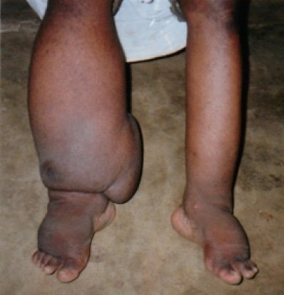
Clinical Manifestations Of Filariasis
Symptoms start after 9 to 12 months, stay in the endemic area. Fever, lymphangitis, lymphadenitis and epididymo-orchitis are early manifestations. Fever is usually moderate and intermittent but sometimes, it may be high and associated with severe rigor, sweating and delirium. Febrile episode, lymphangitis and lymphadenitis usually coincide, but the lymphatic manifestations may preced or follow the fever at times. Inguinal nodes, lower limbs are frequently affected in W. bancrofti infection. In B. malayi infection, genital involvement is rare, and upper limbs are also more frequently involved. Common sites for lymphangitis are the inner aspects of the thighs, legs and arms. In many cases, repeated episodes of lymphangitis lead to distal edema which resolves initially and later on tends to persist. Other common features of the acute episode are lymphedema, funiculitis, orchitis and epididymitis. Less common presentations are scrotal pain, abscess formation in the breast in females, facial lymphangitis, or lymphadenitis in the abdomen resembling a picture of acute appendicitis or psoas abscess. The lymph nodes may become enlarged and spongy (lymphadenovarix). The involved lymphatics may develop abscess which leads to persistent sinuses. Elephantoid lesions develop after repeated acute attacks extending over several years. In some subjects, the acute manifestations recur over long periods, without developing obstructive features, whereas in a few others, the chronic obstructive manifestations develop apparently without the acute episodes.
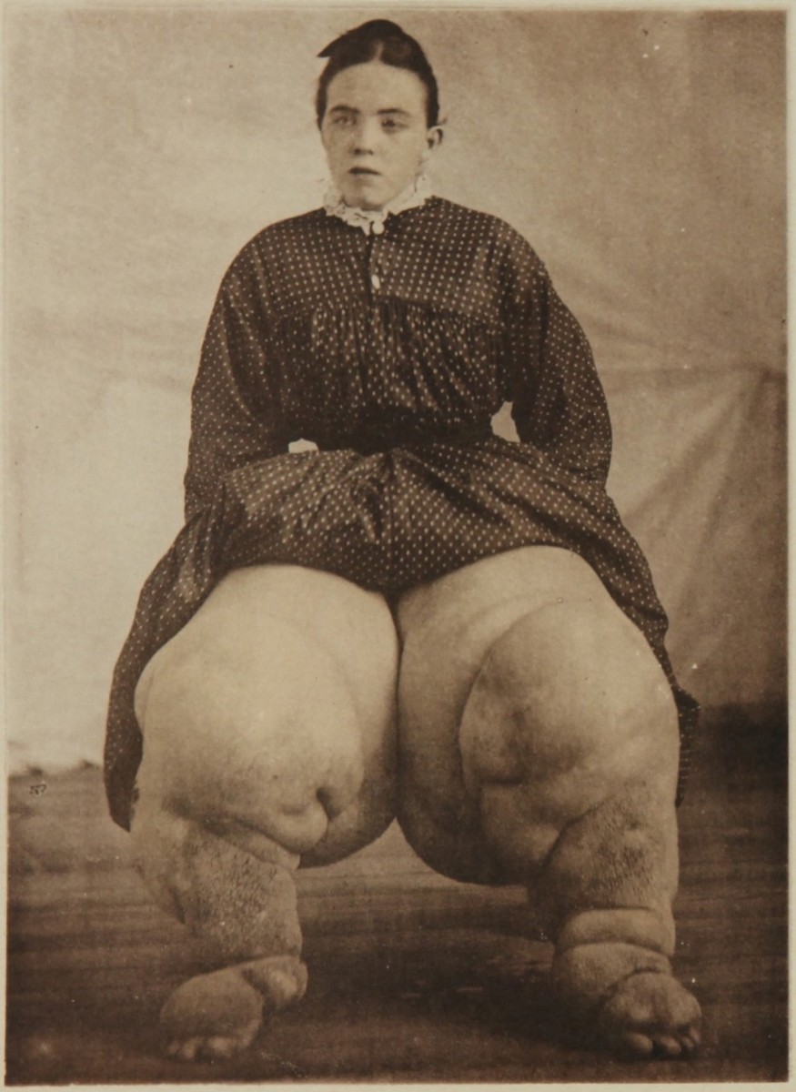
Filariasis Or In Otherwords Known As Elephantiasis
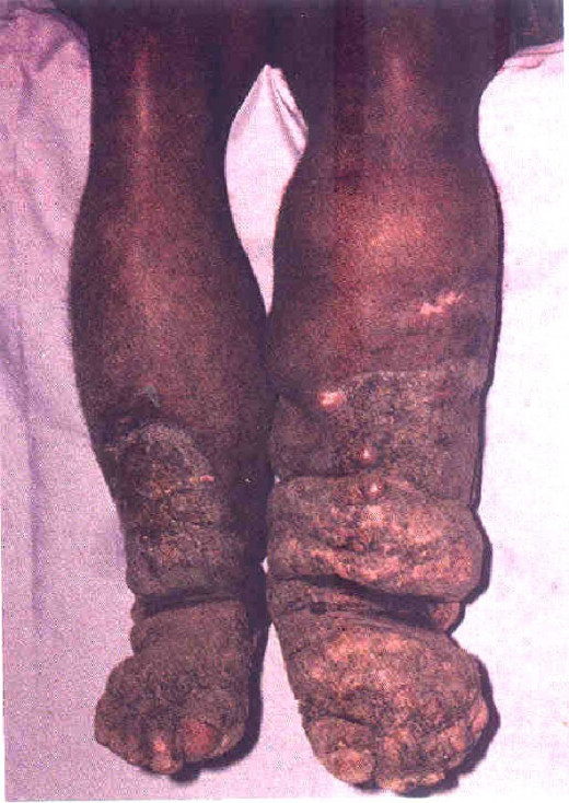
Obstructive Features Of Filariasis
The commonest permanent residuum of the disease is elephantiasis which may attain enormous sizes. Lower limbs, are commonly affected. In females, the breast and the genitalia may also become elephantoid. Hydrocele of the tunica vaginalis which is a common manifestation of the obstructive stage results from involvement of the para-aortic group of lymph nodes. The skin of the scrotum may be covered with vesicles distended with lymph giving a soft velvety fell known as lymph scrotum. Chylous pleural effusions and joint effusion may result from lymphatic obstruction. Chyluria may be due to obstruction of renal or vesical lymphatics. It is often associated with hematuria. Even after entering the chronic obstructive phase, many patients experience acute exacerbation precipitated by factors such as infection, exertion or change of place.
Infectious Diseases
© 2014 Funom Theophilus Makama


