How back pain works? #8 - Spinal Stenosis
INTRODUCTION
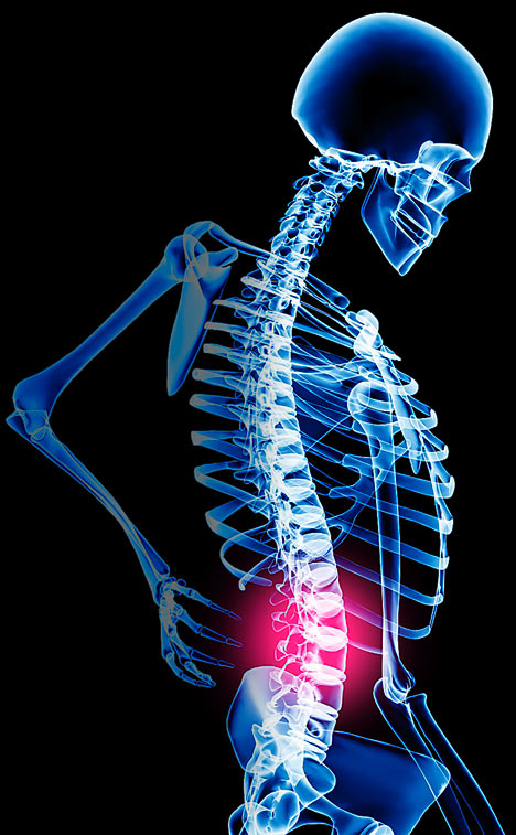
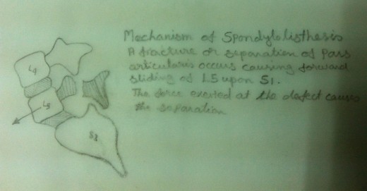
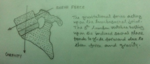
Initially the condition of ‘Spinal Stenosis’ was considered as an anatomical variation that lead to specific neurologic symptoms. Now there is frequent diagnosis of ‘Spinal Stenosis’ ie.Stenosis of one of the canals, either the spinal canal or the intervertebral foramina. Numerous causes are attributed as leading to and causing stenosis. They may be classified into two categories. They are congenital (developmental ) and acquired. The congenital is classified into two sub divisions. ie. idiopathic and achondroplastic. The acquired may be due to Spondylosis (degenerative disk disease) or due to Spondylolisthesis. This condition may also occur after diskectomy or may be due to metabolic bone diseases like Paget’s.
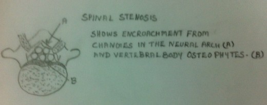
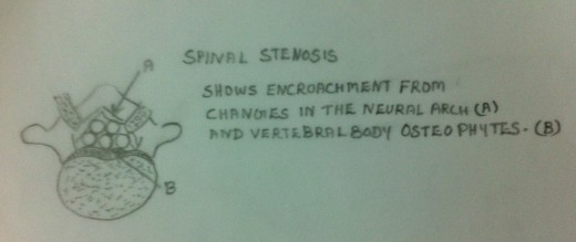
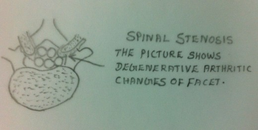
How Spinal Stenosis Occurs
Clinically the Spinal Stenosis is manifested mainly by the presence of paresthesias of one or both legs occurring after a period or distance of walking or after a period of standing. It is also to be noted that in the case of Spinal Stenosis the above mentioned symptoms subside or disappear after sitting or bending forward. In this case the symptoms do not subside or disappear merely by stopping walking.
The compression of the content of the foramina (which is responsible for the symptoms) is from various factors. The factors are;
- In lumbar lordosis the length of the canal is shorter than in lumbar kyphosis.
- The canal is also shortened by disk degeneration at several levels causing the cauda equine to ‘bunch up‘ causing constriction.
- The foramen is also narrowed by anterior osteophytes, posterior exostosis of the foramen or from a hypertrophic superior facet of the inferior vertebra.
- When the lumbar spine is extended the foramen also gets mechanically narrowed.
- While walking there is an increase in venous blood flow (due to dilation) of the veins that accompany the nerve roots as they emerge into the foramina.
- If there is compression of the nerves within the foramina the arterial supply is limited because of the compression caused by the venous return. Hence the symptoms.
The symptoms subside or disappear after sitting or bending forward. This is because of the following factors;
- While sitting flexed forward or standing flexed forward the lumbar lordosis gets changed to lumbar kyphosis.
- The canal lengthens in lumbar kyphosis.
- The caudal equinal fibers elongate.
- The facet separate and the foramina open.
- The venous circulation returns and the blood flow to the nerve returns.
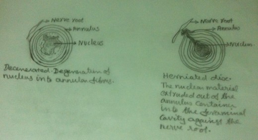
Certain other factors are also responsible for the manifestation of symptoms in spinal stenosis. Standing in one leg causes unequal pressure on the lower discs with compression on the side towards which a person leans. That is, when a person leans to the left the discs gets compressed on the left side. The radius of a normal disc increases by 8% when a person stands on one leg with the lateral bulge of the disc to that side. In the case of a degenerated disc, the radius of the disc may increase by 30% to 35% with the greater bulge within the foramen. While walking or standing, these bulges occur rhythmically into the foramen with our gait. This alternate lateral bulging during walking results in foraminal compression by the disc on the nerve roots. If there is lateral disc bulge, be it an annular bulge from a nuclear extrusion or an extrusion of the nuclear material out of the annulus, this protrusion into the foramina causes ischemia of the nerve roots. Hence the symptoms occur.
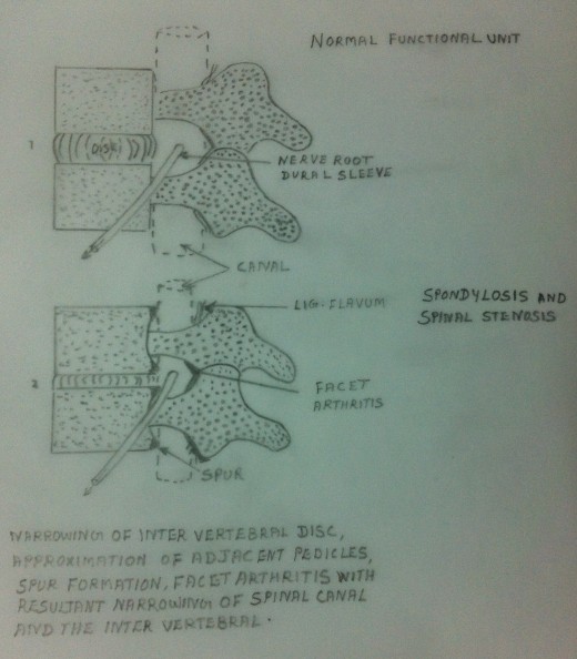
Clinical Manifestations
1.Paraesthesia – presence of paraesthesia of one or both legs occurring after a period or distance of walking or after a period of standing. This symptom subsides or disappears after sitting and bending forward. But this symptom is not relieved merely by stopping walking. The symptoms again recur from a period of walking or standing in a lordotic posture.
2.Neurogenic Bladder Symptoms- occur in spinal stenosis. These symptoms may not have caused any discomfort to the patient. The symptoms include difficulty in maintaining the stream in the standing position (in the case of man). He also feels ease of evacuation in sitting. External abdominal pressure may be needed to completely empty the bladder. The symptoms may also include frequent, urgent and incomplete urination. Dribbling and soiling may be present. Retention of residual urine leads to possible infection with enhancement of the symptoms of frequency and urgency.
3.Confirmation by CT scan, MRI and Myelography- These diagnostic steps assist in determining the type, the extent and the tissues involved.
4. Onset may be tingling, weakness or clumsiness. Pain may be minimal. Often burning sensation or numbness. Saddle distribution of numbness frequent.
Treatment
- Non operative treatment must be undertaken before resorting to surgical intervention. Non operative treatment is preferred when the symptoms are not too severe or not causing progressive organic neurogenic impirement.
- Spinal Extension Exercises must be avoided. Majority of the patients increase the stenosis of the spinal canal with lumbar extension. Hence Spinal Extension Exercises, Position and Posture must be avoided.
- Pelvic Tilt Exercise- In this exercise, the anterior aspect of the pelvis is elevated and the posterior aspect of the pelvis is lowered. Please refer “How to take care of your back” (My earlier article).
- Abdominal Muscle Strengthening Exercises- String abdominal muscles, especially the Oblique Muscles, must be gained and maintained. Please refer “How to take care of your back” (My earlier article).
- Prone Lying must be avoided.
- Spinal Flexion Exercises- Spinal Flexion Exercises are advisable. But prolonged excessive flexion must be avoided as it can cause tension and stretch on the caudal roots. Please refer “How to take care of your back” (My earlier article).
- Low Back Stretching Exercises-Low Back Stretching Exercises are advisable. Avoid exercises that enhance the symptoms. Please refer “How to take care of your back” (My earlier article).
- Hamstring Stretching Exercises- Tight Hamstring Muscles cause an excessive lordosis. Excessive lordosis worsens the symptoms due to spinal stenosis. Hence stretched hamstring muscles must be gained and maintained. Please refer “How to take care of your back” (My earlier article).
- Corset or Brace-A Corset or a Brace may be given. Excessive lordosis worsens the symptoms due to spinal stenosis. Corset or Brace decreases the lordosis and hence minimizes the symptoms. It reinforces the abdominal muscles and minimizes excessive motion of the lumbar spine during lifting or bending. A Corset or Brace must be adjunct to exercise. It must be used along with the exercises. Using the Brace is not to replace the exercise program. Prolonged use of Corset or Brace is not advisable as it tends to diminish the activities of the abdominal muscles and the muscles of the lumbar region. This results in atrophy or loss of flexibility of the abdominal muscles, muscles of the lumbar region and the fascia.
- Electrotherapy Modalities – Short Wave Diathermy (SWD), Ultra Sound Therapy (UST), Transcutaneous Electrical Nerve Stimulator (TENS),Infra Red etc. are also included in the treatment regime for spinal stenosis.
- Pelvic and Gravity Traction- Pelvic and gravity traction are helpful to relieve the symptoms. The traction may be continuous or intermittent.
- Curtailment of walking distances is of very help.
- Evacuation of urine by the patient himself must be taught to him if there are urinary symptoms.
- Oral steroids and Epidural Steroids- may help.
- Surgical decompression- may or may not help.
Prevention of recurrence of back pain due to spinal stenosis is difficult because the problem is anatomically mechanical. Principles of “How to take care of your back” plays an important role in preventing the recurrence of the symptoms due to spinal stenosis. Teaching the patient how the spine normally functions and how the condition of spinal stenosis prevents normal function is very important. Instituting normal and proper body function in everyday activities is very important.

