Measles (Rubeola): Health significance As an Enanthema Infection, Pathology And Clinical Presentations
Rash And Fever In Highly Contagious Measles
A General Overview And Pathology Of Measles
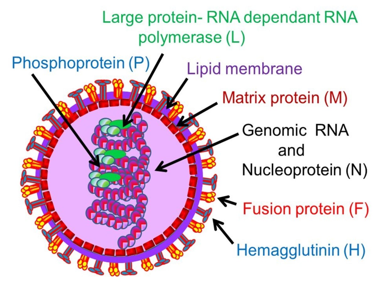
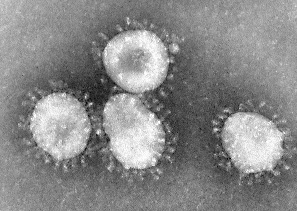
Measles or Rubeola (red spots- Arabic) is an acute exanthematous febrile illness caused by a specific virus of the paramyxovirus group affecting children more. It is an RA virus varying in size from 150 to 300 nm.
Pathogenesis and pathology: Man is the only natural host. The disease may occur in localized or wider outbreaks once in 2 to 3 years, especially during spring. The virus is present in the nasopharyngeal secretions and infection is by droplets. Portals of entry are the respiratory mucous membrane and the conjunctiva. The period of infectivity starts 5 days after the exposure of the virus and lasts until 5 days after skin eruptions have appeared. Infants do not generally suffer, presumably because of the persistence of maternal antibodies in them.
The virus multiplies in the epithelium of the respiratory tract and then causes viremia. Hematogenous spread occurs to various tissues. The virus can be isolated from blood, conjunctiva, lymphoid tissues and secretions of the respiratory tract for a few days before and for 1 or 2 days after the appearance of rashes and from the urine as long as 4 days after the rash.
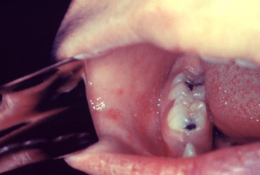
The mucous membrane lesions (enanthem) known as Koplik’s spots seen in the cheeks, consist of vesicles and necrotic epithelium. Microscopically, the lesions show cytoplasmic and intranuclear inclusions, giant cells and intercellular edema. Aggregates of virons in the affected cells can be demonstrated by electron microscopy. Multinucleated giant cells with acidophilic nuclear and cytoplasmic inclusions (Warthin Finkel day cells) may be found in the hyperplastic lymphoid tissues of lymph nodes, tonsils, spleen and thymus. Chromosomal breaks may be seen in leucocytes. Epithelium of the respiratory tract is disrupted and this favours secondary bacterial infection. Lungs show interstitial pneumonia with giant cell infiltration. In patients, who develop encephalomyelitis, the brain shows focal hemorrhage, congestion and perivenous demyelination.
Koplik Spots In Measles
Infectious Diseases
Clinical manifestations Of Measles
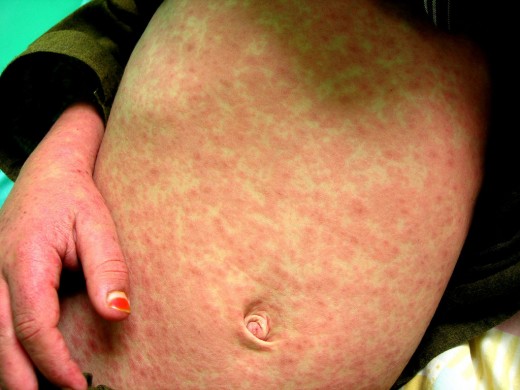
Incubation period is 9 to 11 days. The initial manifestations are malaise, irritability, high fever, conjunctivitis with excessive lacrimation, edema of the eyelids, photophobia, hacking cough and nasal discharge. These symptoms last for 3 to 4 days (up to 8 days) and then the rashes start. Koplik’s spots appear as small, red irregular lesions with bluish-white center on the mucous membrane of mouth 1 to 2 days before the skin rash. They are best seen in the buccal mucosa opposite the upper molar teeth. Lesions similar to them may occur in the conjunctiva and intestinal mucosa. The rashes are red and maculopapular, first, appearing on the fore-head and behind the pinna, later on spreading down to the face, neck, trunk and limbs. Maximum affection is on the face and shoulders. These rashes persist for about 3 days and disappear in the order in which they appeared. The disease becomes more severe in adults with whom complications are likely to be more. The general symptoms disappear 1 to 2 days after the appearance of skin rash. Cough may persist throughout the illness.
© 2014 Funom Theophilus Makama


