Other Rare Forms Of Lymph Node Disorders And All About Bukkitt’s Lymphoma

Clinical Manifestation of Angioimmunoblastic Lymphadenopathy
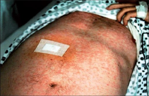
Angioimmunoblastic Lymphadenopathy
This is the result of an immunological disorder seen in patients taking drugs for various conditions. The pathological process suggests a borderline between a reactive condition and a neoplasm.
Histology: The lymph node shows loss of normal architecture, pleomorphic cellular infiltrate consisting of immunoblasts, plasma cells, neutrophils and histiocytes and proliferation of small blood vessels.
Clinical picture: The disease starts as an acute or subacute illness characterized by fever, sweating, weight loss, generalized lymph node enlargement and hepatosplenomegaly. Unlike malignant lymphomas, these patients show a rise in polyclonal immunoglobulins, especially IgM. If left untreated, many cases end fatally due to infective complications or supervening malignancy. The prognosis in indidivual cases is unpredictable.
Treatment is effective in one-third of the cases. Corticosteriods, alkylating agents and combination chemotherapy have all been employed with variable results.
Hydantoin-Linked Lymphoma (Pseudolymphoma)
Diphenul hydantoin may produce an allergic response characterized by fever, rash, generalized lymphadenopathy and even splenomegaly resembling angio-immunoblastic lymphadenopathy. The condition remits fully within months of drug withdrawal.
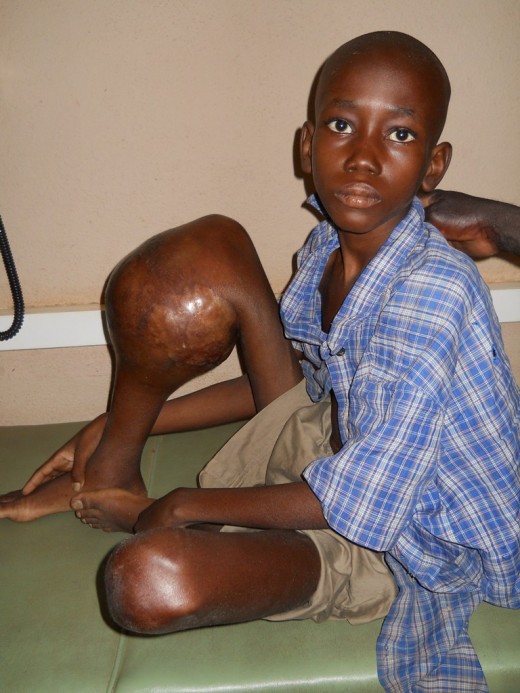
Mycosis Fungoides Knee
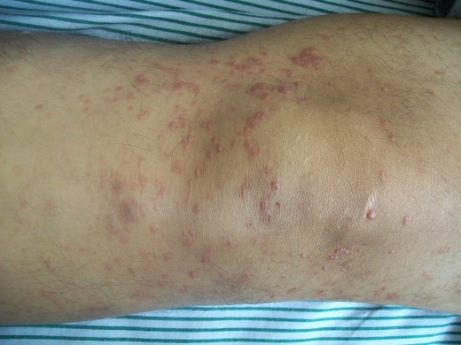

Mycosis Fungoides
This term has been loosely used by different workers. The consensus is to limit the term to the group showing a classical histological picture. Mycosis fungoides and Sezary syndrome are T cell non- Hodgkin’s lymphomas. These are all rare diseases.
Clinical features: The onset of the disease may be as a lesion resembling pruritic erythroderma, psoriasis, seborrheic dermatitis, eczema, nonspecific exfoliative dermatitis, lichenoid dermatitis or neurodermatitis. After several years, plaque formation or tumour formation starts. In some cases, the lesions directly start as tumours which may ulcerate. The skin is red and diffusely thickened. Later the nails may be affected and they may be lost.
The course of the disease is variably. Tumour-like lesions do worse than lichened lesions. Advancing age worsens the prognosis. Median survival period is 5 years from diagnosis.
Clinical Manifestations Of Sezary Syndrome
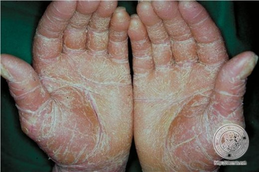
Sezary Syndrome
This is the leukemic phase of mycosis fungoides. It is rare and is characterized by intense pruritis, erythroderma and later on enlargement of the liver, spleen and lymph nodes. Skin shows diffuse infiltration by typical Sezary cells. These cells are seen in good numbers in the peripheral blood.
Treatment: The condition responds to alkylating agents like chlorambucil or cyclophoasphamide singly. Combination therapy as for NHL has been found beneficial in advanced lesions. Superficial irradiation with rays of low penetrance and surface application of mustine hydrochloride (HN2) 10 mg dissolved in 60 ml water brings about remission in a good number of cases. Intensive electron beam therapy or photochemotherapy with Psoralens and long-wave ultraviolet light are newer modalities employed.
Clinical Manifestation Of Bukkitt’s Lymphoma
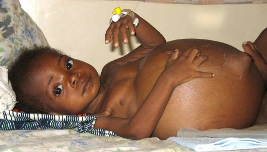
Symptomatology Of Bukkitt’s Lymphoma
Bukkitt’s Lymphoma
This lymphoma is more prevalent in Africa showing a geographical localization to areas with altitude below 1700 meters and high rainfall which are also endemic for malaria. Stray reports are available from all parts of the world, including several from India. Probably, Epstein Barr (EB) virus infection is a cofactor in the development of Burkitt’s Lymphomas (BL), the other factor being chronic stimulation of the reticulo-endothelial system by malaria.
Histology
Histology of the lesion is diagnostic and the tumour is made up of uniform type of lymphoblast-like cells 10- 25 um in size with rounded nuclei and nucleoli. Phagocytic macrophages with large pale cytoplasm are uniformly distributed among the dark staining cells giving the appearance referred to as the “starry sky” appearance.
Clinical features
The African cases are seen predominantly in children, mostly in the age group of 3-5 years. Multicentric tumours arise which grow rapidly affecting the jaw, salivary glands, neck, abdominal viscera, long bones and central nervous system. Spread to bone marrow and lymph nodes and the leukemic phase are incommon.
In the disease, as reported from other parts of the world, older age groups are affected. Lymph node and marrow involvement Is more common. Antibodies to EB virus are less pronounced.
Course and prognosis
The disease is rapidly fatal is untreated. The African cases respond dramatically to cyclophosphamide in large doses or combination chemotherapy. Response is less dramatic in cases described from other parts of the globe.
© 2014 Funom Theophilus Makama


