Post Kala-Azar Dermal Leishmaniasis: Etiology, Pathology, Clinical Presentations, Diagnosis, Treatment And Prognosis
Physical Presentations Of Dermal Leishmaniasis
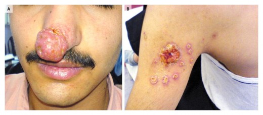
PKDL-Dermal Leishmanoid Of Brahmachari
It is a chronic granuloma of the skin caused by Leishmania donovani following recovery from Kala-azar. Maximum number of cases have been described from India and Bangladesh. In other endemic regions, PKDL is rare or absent.
Etiology: PKDL develops as a sequel in 5 to 10% of cases of Kala-azar, usually is breakdown of cell-mediated immunity in the host so that the parasites in the dermis are most eliminated even after the visceral disease has been cured by chemotherapy or natural resistance.
Pathology: The lesions consist of granulomas in the dermis made up of parasitized macrophages, lymphocytes and plasma cells. The lesions do not ulcerate.
Nodular Lesions Of Cutaneous Leishmaniasis
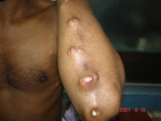
Clinical Features Of Cutaneous Leishmaniasis
Hypopigmented macules: Macular or erythematous lesions develop which are hypopigmented and flush with the surface, varying in size from pin-point to large patches. These may involved the whole body leaving the regions of the belt, axillae and neck unaffected. The face, extensor surface of the limbs and lateral parts of the back show predilection. The whole face and in rare instances the whole body may be affected.
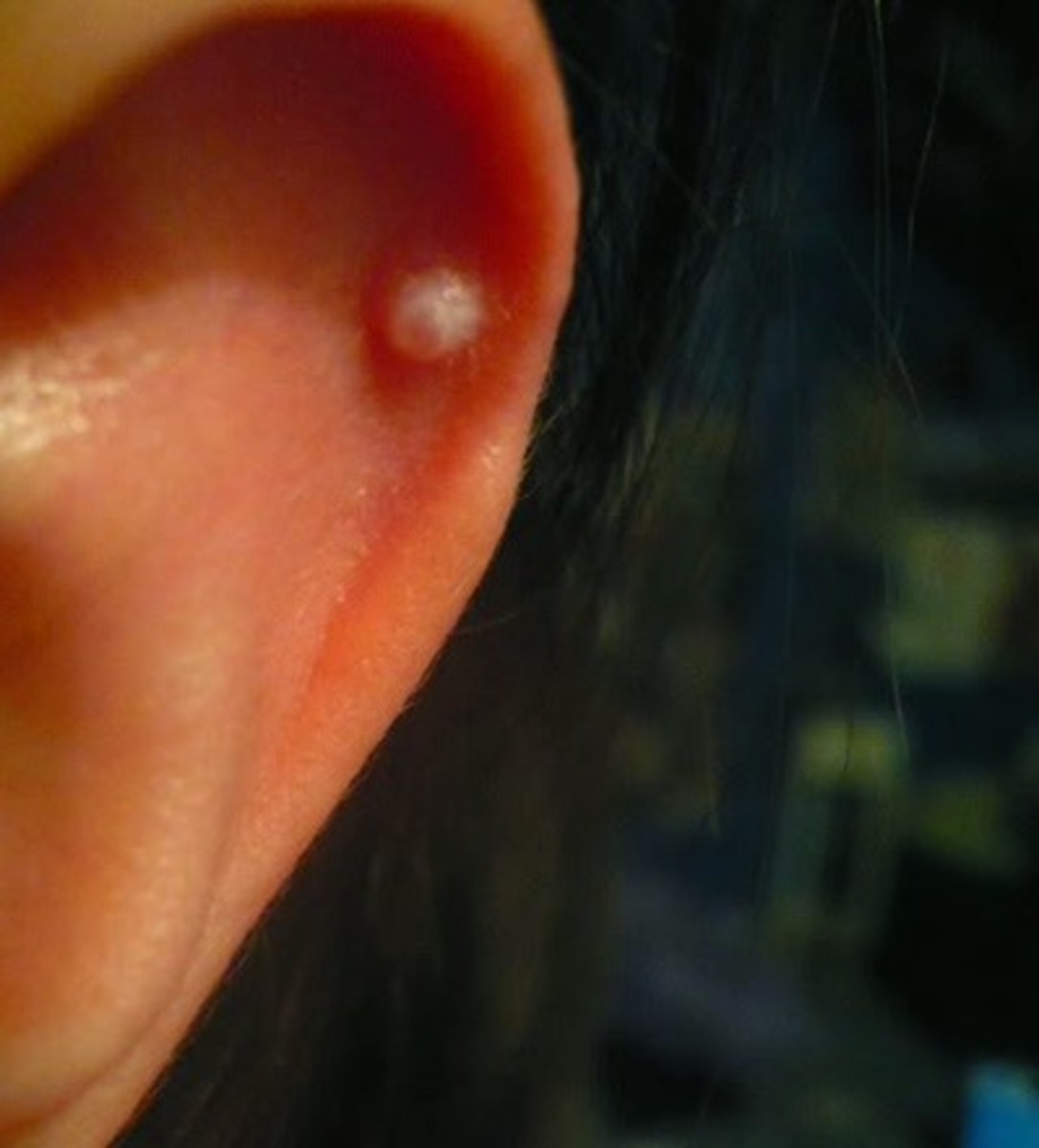
Nodular lesions: They are commonly seen on the chin, other parts of the face except the ears, trunk, limbs, genitals and rarely the mucous membrane of the tongue and lips. Occasionally, large tumour-like masses may appear on the face, limbs and trunk. The majority of cases of PKDL show more than one type of lesion. Constitutional disturbances are not present.
Laboratory features: The aldehyde test is usually negative. Leukocyte levels are normal. The complement fixation test may be positive in a few cases. Indirect hemagglutinin test is usually positive. Serum gammaglobulins (IgG) levels are raised.
Cutaneous Leishmaniasis
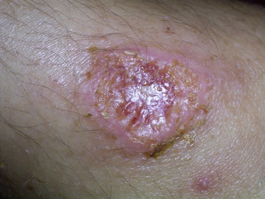
Infectious Diseases
Diagnosis And Treatment Of Cutaneous Leishmaniasis
It is confirmed by demonstrating LD bodies in the “slit and scrape material” obtained from the skin lesions and stained by Leishman stain. Further, L. donovani may be grown in NNN medium from material aspirated from the nodules. The flagellate form (promastigote) is readily demonstrable after 7 to 10 days of incubation. This disorder (PKDL) has to be differentiated from lepromatous leprosy, leucoderma, pityriasis versicolor, dermatitis, lupus erythematosus and neurofibromatosis.
Treatment: It responds to pentavalent antimonials as in the case of Kala-azar. Three to four courses may be required.
Prognosis: If left as such, the lesions progressively worsen and cosmetic deformity results. With treatment, 50% clear completely and others may show considerably improvement. Nodules and erythema readily respond, but the hypopigmented macules are more resistant.
© 2014 Funom Theophilus Makama


