Ray Fungus: Clinical Presentations, Pathology, Prognosis, Treatment And Importance As An Actinomycosis
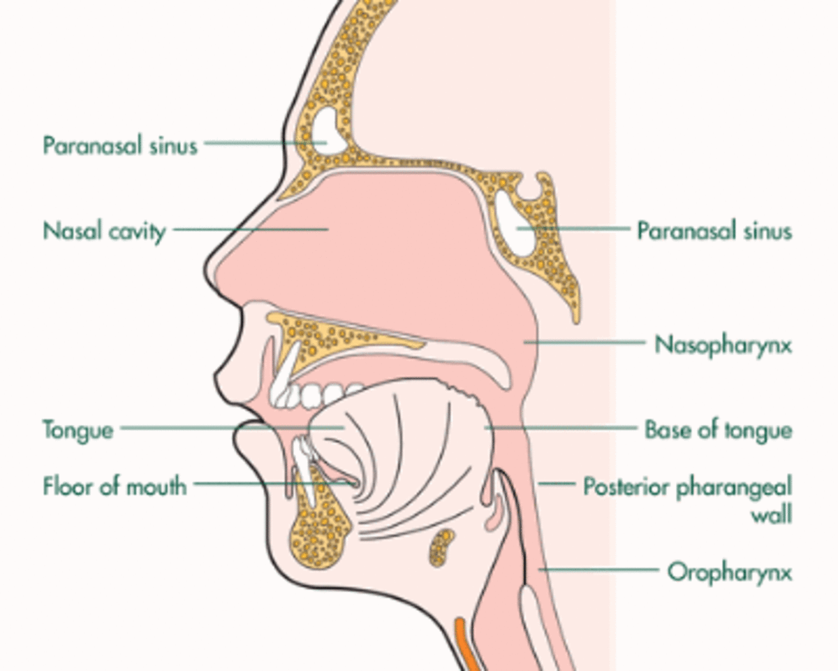
Cervicofacial Form Of Ray Fungal Infection
Actinomycosis
This is a chronic, slow-growing, progressive, granulomatous lesion caused by Actinomyces Israeli. Actinomycetes are transitional forms between bacteria and fungi. They form mycelia like fungi. They are susceptible to antibiotics and in this regard, they behave more like akin to bacteria. They are Gram Positive. There is still controversy about the classification of actinomycetes. The two genera of medical importance are Actinomyces and Nocardia. Actinomyces are anerobic or microaerophilic whereas Nocardia are aerobic. Actinomyces Israeli is present in the mouth as a commensal and tissue invasion occurs due to minor trauma.
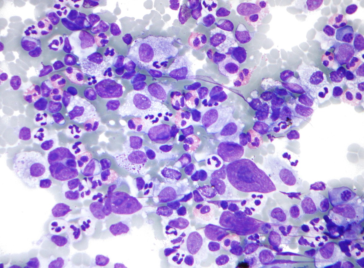
Pathology: The lesions are granulomas with added suppuration. The pus shows the mucelium of the organisms as yellowish granules (sulphur granules). These granules become more distinct when the pus is shaken vigorously with water.
The Thoracic Form Of Ray Fungal Infection

Infectious Diseases
Clinical Manifestation Of Ray Fungus
The disease presents as the cervicofacial form, thoracic form, abdominal form and bony lesions. Symptoms depend on the anatomic location.
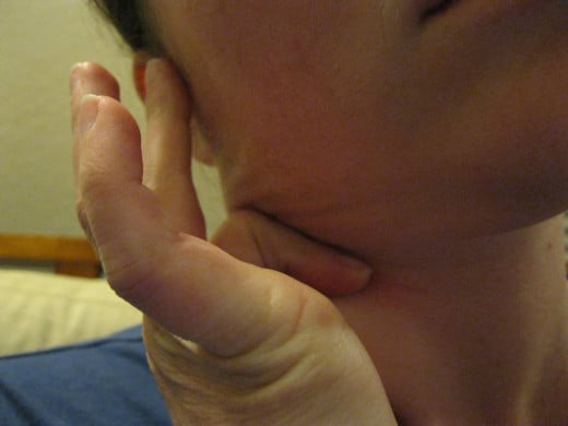
Cervicofacial form: This form starts as a painless, slow-growing lump near the jaw or a painful lesion resembling an abscess. The lesion progresses to involve deeper tissues and bone and sinuses develop. Trismus may occur. Lymph nodes are generally spared. Systemic symptoms are few or absent. The lesion progresses slowly to destroy all tissues. Sinuses discharge the characteristic pus. Presence of extensive destructive lesions in the presence of minimal general symptoms should suggest the possibility of actinomycosis.
Thoracic form: This form results from aspiration of the organism from the mouth. Manifestations include pulmonary infiltration and cavitation, pleurisy, empyema and pericarditis. Pulmonary lesions may be unilateral or bilateral and may closely resemble tuberculosis. The lesion may spread to the pleura and chest wall leading to periosteitis of the ribs and formation of discharging sinuses.
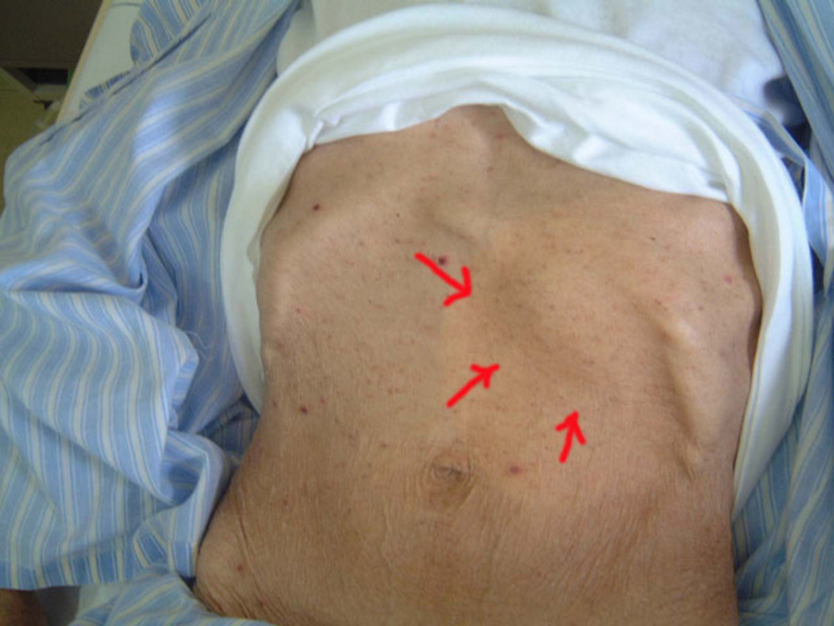
Abdominal form: This form results from ingestion of the organisms. Lesions occur in the cecum and appendix. The disease may present as acute appendicitis of a slow growing lump in the right iliac fossa. This may resemble ileocaecal tuberculosis, Crohn’s disease or malignancy. The lesion may become adherent to the anterior abdominal wall and form sinuses. Rarely the liver and genitals may be involved.
Bone: The bony lesions include osteomyelitis of the mandible, ribs and lumbar vertebrae.
Diagnosis: Strong clinical suspicion is necessary to make the diagnosis. Microscopy of the discharges reveal the mycelia. Histopathological examination confirms the diagnosis.
Prognosis: Early diagnosis and proper treatment result in resolution of the lesions.
Treatment: Penicillin is the drug of choice giving cure rates over 80% in 6 to 8 weeks. Tetracycline and Stilbamidine have to be given when penicillin is contraindicated. Surgery may be necessary in advanced cases.
© 2014 Funom Theophilus Makama


