Salmonella Infections With An Overview On Typhoid Fever, Its Epidemiology And Pathogenesis
The Paratyphoid Pathogens

Salmonella Infections
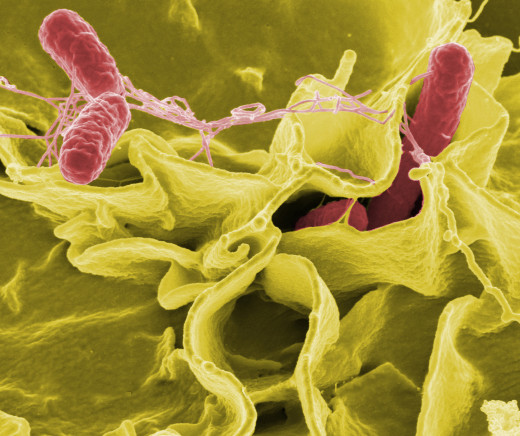
Typhoid and paratyphoid are caused by salmonella typhi and salmonella paratyphi respectively. The genus Salmonella consist of about 1800 serotypes. They are seen in the intestines of vertebrate hosts and are capable of infecting man.
Slamonellae are divided into two groups: the enteric fever group and food poisoning group. Only man is affected by the former group. This includes S. typhi, S. paratyphi A and S. paratyphi B.
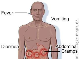
Symptoms Of Salmonellosis

Typhoid Fever
Typhoid fever is an acute febrile disease caused by Salmonella typhi. Salmonella are identified by serologic testing for somatic antigen (O) and flagellar antigen (H). Salmonella typhi itself is known to have more than 1000 species.
Epidemiology: The disease is spread by patients suffering from typhoid fever and the carriers. Persons suffering from enteric fever pass large number of salmonellae in feces and urine. The organisms may be found also in the vomitus, respiratory secretions and other exudates. Carriers may eliminate the organisms in feces (fecal carrier) or urine (urinary carrier).
Salmonella typhi: It can survive for weeks in water, sewage, milk and ice cream. The organisms enter through food and water. Enteric fevers are still common in developing regions, though this infection has been effectively controlled in the developed countries.
Pathogenesis: The incubation period is usually 10 to 14 days. About 107 bacilli are required to cause infection. The factors that determine the development of lesions are the virulence of the organism, size of the inoculums, gastric acidity and the bacterial flora of the upper small intestine. The bacilli enter through the intestinal epithelial lining of the jejunum and ileum and reach the submucosa where they are phagocytosed by the polymorphs and macrophages. The organism survive within the phagocytes and they reach the mesenteric lymph nodes. There, they multiple and enter the blood stream to produce a transient bacteremia. The bacilli reach the liver, gall bladder, spleen, bone marrow, lymph nodes, lungs and kidneys where further multiplication occurs. Large number of bacilli are discharged from the gall bladder into the intestines. This time, they enter the Peyer’s patches and lymphoid follicles of the ileum. These lymphoid structures undergo inflammation, necrosis and ulceration. Clinical manifestations start with the bacteria begin to re-enter the blood stream from the intracellular sites. The infection of the gall bladder is usually asymptomatic. The organism produces endotoxin which accounts for the fever and many of the systemic effects.
Pathology: Spleen and other lymphoid tissues show proliferation of large mononuclear cells and formation of ill-defined nodular collections of reticuloendothelial elements. The liver is enlarged and shows cloudy swelling. The peyer’s patches become swollen and show infiltration by large number of mononuclear cells with ingested S.typhi. Surface of the peyer’s patches undergoes necrosis and sloughing. When the slough separates, ulcers are found in the terminal portion of the ileum. Blood vessels may be eroded and this may result in intestinal hemorrhage. The ulceration may extend to the muscularis mucosa and serosa and result in intestinal perforation.
The cardiac muscle shows areas of degeneration and focal necrosis. Bronchitis and pneumonia may develop. Other tissues may be affected; important lesions being meningitis, osteomyelitis, cholecystitis, cholelithiasis and zenker’s degeneration of muscles, especially the rectus abdominis.
© 2014 Funom Theophilus Makama


