Schistosomiasis: Epidemiology, Pathology, Usual Patient Complaints And Clinical Manifestations
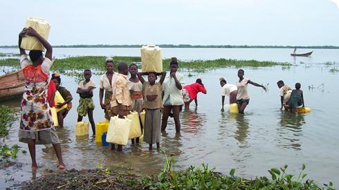
Schistosomiasis Prevails In Mostly Subsahara Africa And Also Other Developing Countries
Epidemiology Of Schistosomiasis
Though schistosomiasis is one of the more widespread helminthic infections affecting about 200 million people in 71 countries, it is only rarely reported from India. There is a pocket of infection by S. hematobium in Maharashtra state. Highest prevalence is in Africa and the Middle East. Schistosoma hematobium and S. mansoni occurs in Africa, Middle East and South America. Schistosoma japonicum is prevalent among the field workers in China, Japan and Philippines, Thailand, Lagos, Vietnam and Burma. Newer areas have become endemic with the construction of dams and irrigation projects.
Pathology and pathogenesis: Eggs are laid in the tissues, but many are diacharged into the lumen and are passed in urine or feces. They are not harmful to the host. About 50% of the eggs are retained in the tissues and they cause granulomatous reactions. Finally, fibrosis occurs. Granulomatous reactions. Finally, fibrosis occurs. Granulomatous elsions occur especially in liver, intestines, urinary bladder, uterus and lungs, resulting in complications. The tissue reaction is determined by the intensity of infection and the reactivity of the host. Ultimately, further reinfection is limited by immunity developed by the host in endemic areas.
Late Complications Of Schistosomiasis

Infectious Diseases
Clinical features and complications Of Schistosomiasis
Three phases are recognizeable:
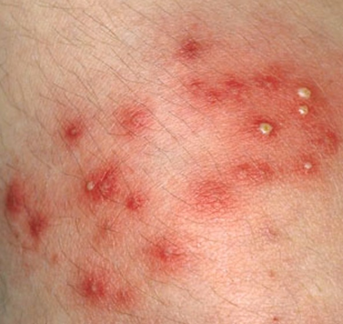
- Earliest lesion is caused by penetration of the skin by cercariae. It results in itching and papular rashes at the sites of penetration (swimmer’s itch, cercarial dermatitis).
- In about 4 to 6 weeks, the second stage starts with fever, severe toxemia and allergic reactions. Marked eosinophilia develops in about 4 to 6 weeks. This stage is called Katayama syndrome. This serum sickness like reaction is seen during ovipositio and it is most severe in S. japonicum infection.
- Late complications are seen three months to several years after the initial infection. These are due to granulomatous lesions and fibrosis occurring in various organs. The manifestations of this stage differ in the three species.
Schistosoma hematobium (Genito-urinary schistosomiasis): affects the bladder, uterus and the prostate in males, whereas vagina, cervix and uterus are also involved in females. Urinary symptoms predominate depending on the stage of the disease. Schistosoma hematobium infection predisposes to vesical carcinoma.
Schistosoma mansoni infection (Intestinal Schistosomiasis): The early stage is characterized by abdominal pain and dysenteric symptoms. The lesion extends to the liver due to retrograde passage of eggs into the portal system and the liver. Periportal fibrosis resulting in presinusoidal portal hypertension develops in the liver. Massive splenomegaly may occur. The ova may reach all organs through the portal-systemic collaterals. The ova in the brain lead to neurological symptoms. Schitosoma mansoni infection favours the development of carrier state for salmonella typhi. There is no rise in the risk of cancer.
Schistosoma japonicum infection (Katayama disease Asiatic schistosomiasis): Since this worm produces more eggs than the others, its pathogenicity is greater. Major pathological lesions are seen in the small intestine, mesentery and ascending colon. This leads to ulcerations, fibrous thickening and polyp formation. Early symptoms are abdominal pain and bloody mucoid stools. Cirrhosis liver develops with all its attendant complications. 5% of cases develop ectopic foci of infection, especially in the lungs and central nervous system. Symptoms include Jacksonian epilepsy, focal paralysis, paraplegia and coma. Embolisation of eggs in the pulmonary circulation leads to obstruction of small arterioles resulting in pulmonary hypertension. Granulomas develop in the lungs which result in fibrosis. Such patients present with asthma, bronchitis or pulmonary emphysema.
© 2014 Funom Theophilus Makama


