Staphylococcal Infections: Health Effects Of Its Various Clinical Presentations As Superficial Staphylococcal Lesions
A Patient Suffering From Staphylococcal Infection

Superficial Staphylococcal Lesions
Superficial lesions are typical of staphylococcal infections and this is aids a lot in their diagnosis. Clinical presentations such as furuncle, carbuncle, impetigo, ecthyma, sycosis barbae, follicular impetigo of Bockhart, scalded skin syndrome, staphylococci pneumonia, bacteremia and osteomyelitis are typical manifestations.
Superficial Staphylococcal Lesions
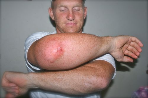
Furuncle: A furuncle is an acute necrotic infection of a hair follicle. It presents as a small follicular inflammatory nodule, soon becoming pustular and then necrotic. It heals after discharge of the necrotic core to leave a violaceous macule and ultimately a permanent scar.
Carbuncle: It is a large furuncle or an aggregate of interconnected furuncles accompanies by intense inflammatory changed in the surrounding and underlying connective tissue. Sites of predilection are the back of the neck, the hips and thighs. It starts as a hard red lump, at first smooth and acutely tender. It slowly increases in diameter to reach 3 to 10 cm. Suppuration begins after 5 to 7 days and pus is discharged from multiple follicular orifices. Constitutional symptoms are usually severe. Carbuncles are particularly common in diabetics and those with lowered general resistance.
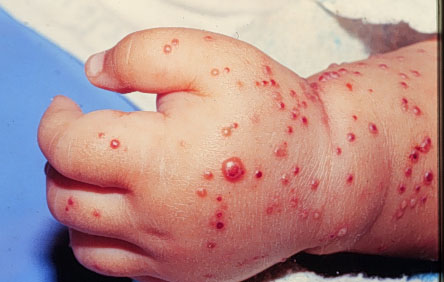
Impetigo: Bullous impetigo is purely staphylococcal. It usually occurs in new- borns and infants. Face is more often affected although, lesions may occur in the palms and soles. The lesions consist of multiple pustules.
Ecthyma: Both staphylococci and streptococci can cause ecthyma. The lesion is characterized by small bullae or pustules on an erythematous base, soon developing into a hard crust. Irregular ulceration is evident when the crust is removed. The sites of predilection are the buttocks, thighs and legs.
Sycosis barbae: This is seen in males after puberty and the lesions are seen on the beard region. The lesion starts as an edematous, red, follicular papule or pustule, centered by a hair. The individual papules remain discrete.
Follicular impetigo of Bockhart: This is staphylococcal infection of the hair follicle seen commonly in childhood. It occurs mainly in the scalp or scalp margins.
The scalded skin syndrome: Also known as pemphigus neonatorum, Ritter’s disease and toxic epidermal necrolysis. This is a generalized exfoliative dermatitis caused by staphylococcus aureus. It is characterized by generalized painful erythema and large bullae which can be easily disrupted by finger- pressure. Newborns are commonly affected.
Skin Lesions In Staphylococcal Infections

Pneumonia And Bacteremia
Staphylococcal Pneumonia: Staphylococcal pneumonia occurs as a complication of influenza, measles or other viral infections. It starts with high fever, chills, dyspnea, cyanosis, cough and pleural pain. Sputum may be bloody or purulent. Scattered fine to coarse rales and rhonchi may be heard over the involved areas. Typical consolidation is rare. Primary staphylococcal pneumonia may occur in infants and young children and this causes pneumothorax, pneumotoceles or empyema. Hematogenous secondary staphylococcal pneumonia is frequent in drug addicts who also develop endocarditis. Staphylococcal pneumonia carries a higher mortality, especially in the old and debilitated patients.
Osteomyelitis: Majority of cases of primary osteomyelitis are due to staphylococcus.
Staphylococcal bacteremia: The primary source consists of local staphylococcal infections, urinary catheters, foreign bodies, or infected intravenous shunts. The onset is marked by high fever, tachycardia, cyanosis and vascular collapse. Pyemic abscesses may form in the skin, bone, kidneys, brain, or lungs. Fulminant sepsis may lead to death within 24 hours.
Other staphylococcal lesions include endocarditis, pericarditis, arthritis, pyomyositis, parotitis and urinary tract infection.
In conclusion, the clinical diagnosis should be confirmed by culturing the organism from the exudates. Phage typing is employed for identifying the strains in epidemics.
© 2014 Funom Theophilus Makama


