Intro to Anatomy 3 (BRAIN/NERVES)

An Introduction to Anatomy of the Human Body
This is a continuation of the "Introduction to Anatomy" series of websites I have created which address different areas of basic anatomy. This lens is looking at the nervous system and brain specifically. I will discuss the function, location, type and name of the cranial nerves as well as where cranial nerves leave the skull. I also describe the lobes of the brain and the function of each. I have also included a detailed diagram of the bracial plexius nerves.
It is a work in progress and will be updated with new material soon.
See my other lenses
Thank you very much for your support!!!
---Learn more about that brain!!!---

Layers of the SKULL and MENINGES
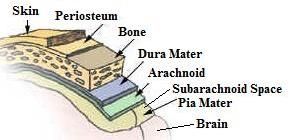
-Skin (epidermis, dermis, subcutaneous) Layers of the skin
-Periosteum The covering that provides the innervation and blood supply to the bone.
-Bone Hard material that is designed to be protective and a structural framework.
-Dura Mater: Periosteal The outer layer that makes contact with the skull.
-Dura Mater: Meningeal The inner layer that makes up the outer sinus walls.
-Venous Sinuses This is where CSF joins deoxygenated blood to return to the heart.
-Arachnoid a thicker membrane layer.
-Arachnoid Space This space is filled with cerebral spinal fluid (CSF) which provides nutrients, waste disposal and shock absorption for the brain.
-Pia Mater (translates to "soft mother") This is the very thin membrane sack surrounding the brain.
BRAIN Lobes
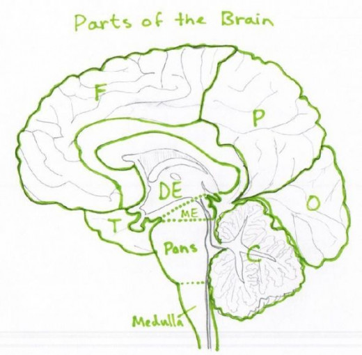
Frontal Lobe (F): Center for higher functioning.
Parietal Lobe (P): Integration of Sensory input.
Temporal Lobe (T): Auditory and Speech Center.
Occipital Lobe (O): Visual processing Center.
Also shown in the picture above are the:
-Diencephalon (DE),
-Mesencephalon (ME),
-Pons,
-Medulla Oblongata,
-Cerebellum(C))
NERVE locations inside the skull

Here are the pathways for each cranial nerve to leave (and sometimes enter) the skull.
Brain and Nerves Resources
- Brain and Meninges
Nice short descriptions and pictures - Nervous System
Details and pictures of the nervous system. - Cranial Nerves
Location of each nerve and clinical application of lesions to that region. - Anatomy of the Brain
A list of structures of the brain and function. - Facial Nerves
What they do, where they go and how they get there. - CSF and the Ventricles
An overview of the Cerebral spinal fluid system, its function, location and clinical application. - Brachial Plexus Tutorial (Cadaver)
A wonderful dissection of the brachial plexus in a cadaver with each structure pointed out and labeled in a video. - Eye problems
Common deficits of the eye are explained. - Cranial Nerves Attachments
A picture showing where they attach to the brain.
Cranial Nerves - Nerve Name, Nerve Type, Nerve Location and Nerve Function
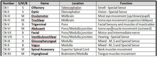
* NOTES*
-S/M/B are the type of nerve (Sensory is S, Motor is M and Both is B)
-Trigeminal nerve splits into the Opthalamic (1), Maxillary (2) and Mandibular (3) branches
---Brain and Nerves Shop---


NERVES of the Brachial Plexus

1) Dorsalscapular: innervates the Rhomboids, Levator scapulea
2) Suprascapular: Supraspinatus, Infraspinatus
3) Subclavian: Subclavius
4) Lateral Pectoral: Pectoralis Major
5) Musculocutaneous: upper arm flexors (Biceps Brachii, Coracobrachialis, Brachialis)
------------------------------------------------------------------------------------------------------------
6) Upper Subscapular: Subscapularis
7) Thoracodorsal: Latissimus Dorsi
8) Lower subscapular: Subscapularis, Teres Minor
9) Radial: Posterior Arm/Forearm (Triceps, Anconeus, Brachioradialis, Supinator)
10) Axillary: Deltoids, Teres Minor
-------------------------------------------------------------------------------------------------------------
11) Medial Pectoral:Pectoralis Major, Pectoralis Minor
12) Medial Brachial Cutaneous: sensory nerve of the skin
13) Ulnar: Flexor carpi ulnaris, frexor digitorum profundus, hand muscles
-------------------------------------------------------------------------------------------------------------
14) Long Thoracic: (5,6,7 Serratus Anterior)
15) Phrenic: (3,4,5 keep the diaphram alive)
16) Median: Flexor carpi radialis, flexor digitorum superficialis, flexor digitorum profundus, flexor pollicis longus
1 - 5 : nerves spliting from the most superior and lateral continuous nerve
6 - 10: nerves spliting from the deepest and most center continuous nerve.
11 - 13: nerves spliting from the most inferior and medial continuous nerve
14 - 15: nerves spliting from multiple rami
16:The nerve that is formed form the most lateral and most medial continuous nerves
*CADAVER- Brachial Plexus Tutorial*
WARNING- A REAL HUMAN CADAVER IS USED FOR THIS DISSECTION. IF YOU ARE SQUEAMISH I SUGGEST NOT VIEWING THE VIDEO BELOW.
A dissection of the brachial plexus where each section is clearly described and shown on a human cadaver. A very helpful resource for locating the nerves of the brachial plexus.
The Cranial Nerves Song
Digestive System Resources
- Digestive System
Pictures and descriptions of structures (mouth, esophagus, stomach, small and large intestine and bile system. - Stomach
Pictures and descriptions of the stomach, arteries, veins and nerves of the area. - Liver
Lots of good pictures and description of the liver and bile system.


